 Back
BackThe Central Nervous System: Structure and Function
Study Guide - Smart Notes
 Tailored notes based on your materials, expanded with key definitions, examples, and context.
The Central Nervous System (CNS)
Overview of the CNS
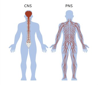
The central nervous system (CNS) is composed of the brain and spinal cord. It serves as the main control center for processing and integrating information in the body, coordinating sensory input and motor output.
Brain: Responsible for higher functions such as thought, memory, emotion, and voluntary movement.
Spinal Cord: Conducts signals to and from the brain and controls reflex activities.
Embryonic Development of the Human Brain
Neural Tube and Brain Vesicles
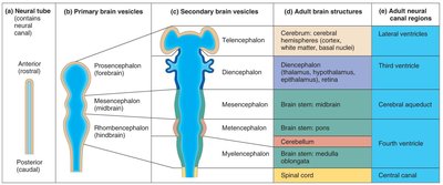
The human brain develops from the neural tube, which forms three primary brain vesicles: the prosencephalon (forebrain), mesencephalon (midbrain), and rhombencephalon (hindbrain). These vesicles further differentiate into secondary brain vesicles and adult brain structures.
Primary Vesicle | Secondary Vesicle | Adult Structure | Neural Canal Region |
|---|---|---|---|
Prosencephalon | Telencephalon | Cerebrum (cerebral hemispheres, cortex, white matter, basal nuclei) | Lateral ventricles |
Prosencephalon | Diencephalon | Thalamus, hypothalamus, epithalamus, retina | Third ventricle |
Mesencephalon | Mesencephalon | Midbrain (part of brain stem) | Cerebral aqueduct |
Rhombencephalon | Metencephalon | Pons, cerebellum | Fourth ventricle |
Rhombencephalon | Myelencephalon | Medulla oblongata (part of brain stem) | Fourth ventricle |
Spinal cord | - | Spinal cord | Central canal |
Brain Flexures and Growth
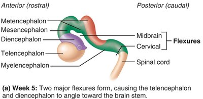
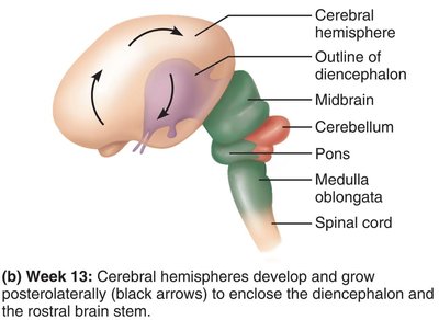
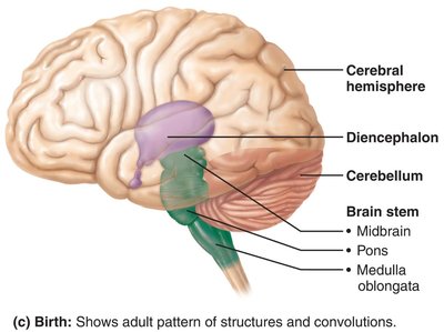
During development, the brain grows faster than the skull, causing it to fold and form flexures. The telencephalon and diencephalon angle toward the brain stem, and the cerebral hemispheres envelop the diencephalon and midbrain.
Pattern of Distribution of Gray and White Matter in the CNS
Gray Matter vs. White Matter
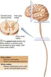
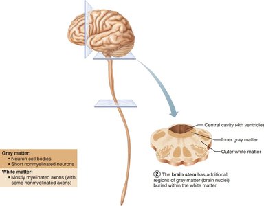
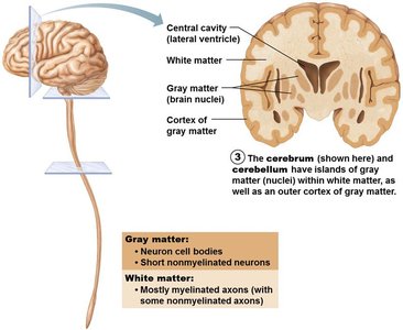
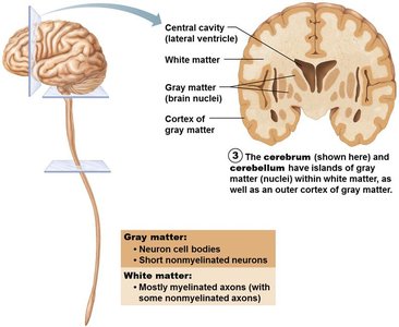
Gray matter consists mainly of neuron cell bodies and nonmyelinated neurons, while white matter is composed primarily of myelinated axons. The arrangement of gray and white matter varies in the spinal cord, brain stem, and cerebrum/cerebellum.
Spinal Cord: Central cavity surrounded by gray matter, with outer white matter.
Brain Stem: Similar to spinal cord but with additional gray matter nuclei within white matter.
Cerebrum and Cerebellum: Islands of gray matter (nuclei) within white matter, plus an outer cortex of gray matter.
Ventricles of the Brain
Structure and Function
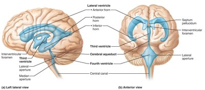
The brain contains four fluid-filled ventricles that are continuous with each other and the central canal of the spinal cord. They are filled with cerebrospinal fluid (CSF) and lined by ependymal cells, which are a type of neuroglia.
Lateral Ventricles: Paired, separated by the septum pellucidum, connected to the third ventricle via the interventricular foramen.
Third Ventricle: Located in the diencephalon, connected to the fourth ventricle via the cerebral aqueduct.
Fourth Ventricle: Located in the hindbrain, continuous with the central canal of the spinal cord.
Cerebral Hemispheres
Surface Features and Lobes
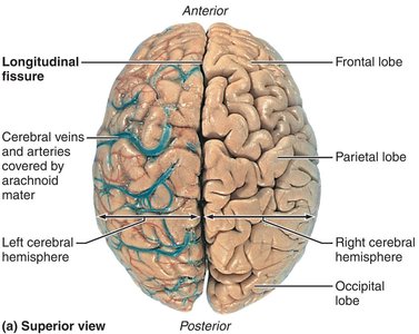
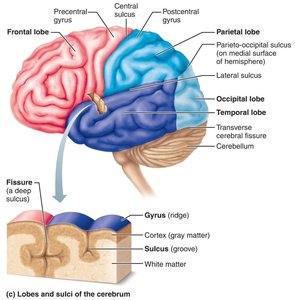
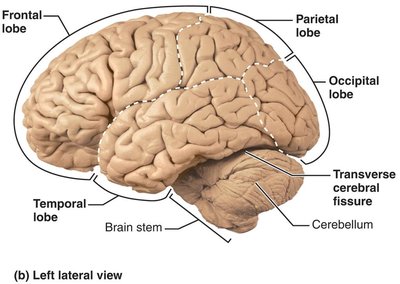
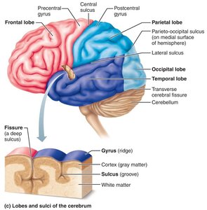
The cerebral hemispheres form the superior part of the brain and account for about 83% of its mass. The surface is marked by gyri (ridges), sulci (shallow grooves), and fissures (deep grooves). Major fissures include the longitudinal fissure (separates hemispheres) and the transverse cerebral fissure (separates cerebrum from cerebellum).
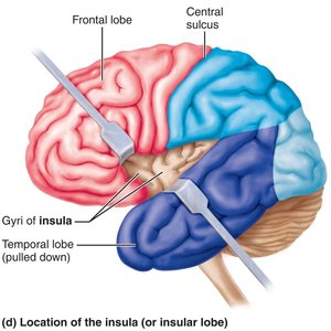
Lobes: Each hemisphere is divided into five lobes: frontal, parietal, temporal, occipital, and insula (buried under portions of the other lobes).
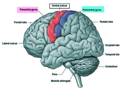
Major Sulci: Central sulcus (separates frontal and parietal lobes), parieto-occipital sulcus, and lateral sulcus (outlines temporal lobe).
Internal Organization
Each hemisphere has three basic regions:
Cerebral Cortex: Superficial gray matter, site of conscious mind.
White Matter: Internal, composed of myelinated axons.
Basal Nuclei: Deep within white matter, involved in motor control.
Cerebral Cortex
Structure and Function
The cerebral cortex is the executive suite of the brain, responsible for conscious thought, perception, and voluntary movement. It is a thin layer of gray matter (about 2–4 mm thick) containing neuron cell bodies, dendrites, glial cells, and blood vessels, but no axons. It makes up about 40% of the brain's mass.
Functional Areas of the Cortex
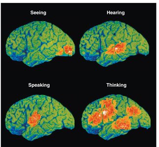
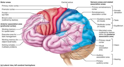
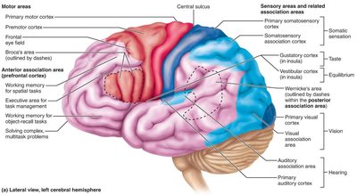
Motor Areas: Control voluntary movement (located in the frontal lobe).
Sensory Areas: Provide conscious awareness of sensation (located in parietal, insular, temporal, and occipital lobes).
Association Areas: Integrate diverse information for purposeful action.
Each hemisphere controls the opposite side of the body (contralateral control), and there is lateralization (specialization) of cortical functions.
Motor Areas of the Cortex
Primary Motor Cortex
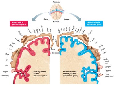
Located in the precentral gyrus of the frontal lobe, the primary motor cortex contains large pyramidal cells whose axons form the pyramidal (corticospinal) tracts. It exhibits somatotopy, meaning specific regions control specific muscles.
Damage: Results in paralysis of voluntary muscles controlled by the affected area.
Premotor Cortex
Located anterior to the precentral gyrus, the premotor cortex helps plan movements, especially complex, learned, or patterned motor skills. It coordinates voluntary actions that depend on sensory feedback.
Damage: Loss of programmed motor skills, but not individual muscle strength.
Broca’s Area and Frontal Eye Field
Broca’s Area: Usually present in the left hemisphere; involved in speech production and planning motor activities.
Frontal Eye Field: Controls voluntary eye movements.
Sensory Areas of the Cortex
Overview
Sensory areas are responsible for conscious awareness of sensation and are located in the parietal, insular, temporal, and occipital lobes. Major sensory areas include:
Primary somatosensory cortex
Somatosensory association cortex
Visual areas
Auditory areas
Vestibular cortex
Olfactory cortex
Gustatory cortex
Visceral sensory area
Primary Somatosensory Cortex
Located in the postcentral gyri of the parietal lobe, this area receives information from sensory receptors in the skin and proprioceptors in muscles, joints, and tendons. It is capable of spatial discrimination, identifying the body region being stimulated.
Somatosensory Association Cortex
Located posterior to the primary somatosensory cortex, it integrates sensory input for understanding objects. Damage impairs the ability to recognize objects by touch.
Visual and Auditory Areas
Primary Visual Cortex: Located in the occipital lobe; receives visual information from the retinas.
Visual Association Area: Surrounds the primary visual cortex; interprets visual stimuli using past experiences.
Primary Auditory Cortex: Located in the temporal lobe; interprets information from the inner ear.
Auditory Association Area: Posterior to the primary auditory cortex; stores memories of sounds and permits perception of sound stimuli.
Other Sensory Areas
Vestibular Cortex: Responsible for conscious awareness of balance; located in the posterior insula and adjacent parietal cortex.
Olfactory Cortex: Located in the temporal lobe; involved in conscious awareness of odors.
Gustatory Cortex: Located in the insula; involved in taste perception.
Visceral Sensory Area: Posterior to the gustatory cortex; conscious perception of visceral sensations (e.g., upset stomach, full bladder).
Association Areas of the Cortex
Integration and Higher Functions
Association areas receive input from multiple sensory areas and send outputs to multiple areas. They are involved in integrating information for purposeful action and higher cognitive functions. The general flow of information is: sensory receptor → primary sensory cortex → sensory association cortex → multimodal association cortex.
Anterior Association Area (Prefrontal Cortex): Involved in intellect, cognition, recall, and personality. Development depends on social feedback.
Posterior Association Area: Involved in recognizing patterns and faces, localizing the body in space, and understanding language (Wernicke’s area).
Limbic Association Area: Provides emotional impact and helps establish memories; includes the cingulate gyrus, parahippocampal gyrus, and hippocampus.
Lateralization of Cortical Function
Specialization of Hemispheres
Lateralization refers to the division of labor between the two cerebral hemispheres. Although both hemispheres share many functions, each is specialized for certain tasks.
Left Hemisphere: Dominant for language, math, and logic.
Right Hemisphere: Dominant for visual-spatial skills, intuition, emotion, and artistic/musical abilities.
Hemispheres communicate via fiber tracts for functional integration.
