 Back
The Central Nervous System: Structure, Function, and Integration
Study Guide - Smart Notes
 Tailored notes based on your materials, expanded with key definitions, examples, and context.
The Central Nervous System (CNS)
Overview of CNS Functions
The central nervous system (CNS) is responsible for integrating sensory information, making decisions, and coordinating motor output. It consists of the brain and spinal cord, which work together to maintain homeostasis, process sensory input, and generate appropriate responses.
Sensory Functions: Detection of sensations inside and outside the body.
Integrative Functions: Decision-making processes, including interpretation and integration of sensory input.
Motor Functions: Stimulation of muscle contractions and glandular secretions.
While sensory and motor functions are performed by the peripheral nervous system (PNS), integrative functions are exclusive to the CNS.
Basic Structure of the Brain and Spinal Cord
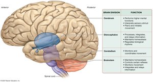
The brain is a soft, whitish-gray organ located in the cranial cavity, composed primarily of nervous tissue. It contains four main divisions and internal cavities called ventricles, which are filled with cerebrospinal fluid (CSF) for protection and support. The spinal cord is a long, tubular structure encased within the vertebral cavity, serving as a conduit for information between the brain and the rest of the body.
Brain Weight: 1,250–1,450 g; receives about 20% of total blood flow at rest.
Spinal Cord: Begins at the foramen magnum and ends between the first and second lumbar vertebrae; central canal filled with CSF.
White Matter and Gray Matter
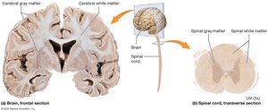
White matter consists of myelinated axons, while gray matter is composed of neuron cell bodies, dendrites, and unmyelinated axons. In the brain, gray matter forms the outer cortex and is scattered in deeper regions, while in the spinal cord, gray matter is internal and white matter is superficial.
Development of the CNS
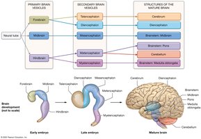
The CNS develops from a neural tube, which forms primary brain vesicles (forebrain, midbrain, hindbrain) that further differentiate into secondary vesicles and mature brain structures.
Forebrain: Telencephalon (cerebral hemispheres), Diencephalon
Midbrain: Mesencephalon
Hindbrain: Metencephalon (cerebellum, pons), Myelencephalon (medulla oblongata)
The Cerebrum
Structure and Lobes
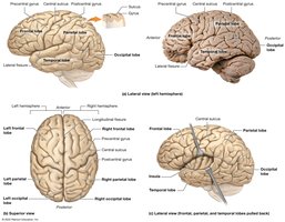
The cerebrum is the largest part of the brain, divided into right and left hemispheres. Its surface is marked by gyri (ridges), sulci (shallow grooves), and fissures (deep grooves). The major lobes are:
Frontal Lobe: Involved in voluntary movement, planning, and higher cognitive functions.
Parietal Lobe: Processes sensory information related to touch, temperature, and proprioception.
Temporal Lobe: Involved in auditory processing and memory.
Occipital Lobe: Responsible for visual processing.
Insula: Deep within the lateral fissure, involved in taste and visceral sensation.
Cerebral Cortex and Functional Areas
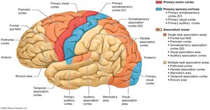
The cerebral cortex, particularly the neocortex, is responsible for conscious processes. It contains primary motor and sensory cortices, as well as association areas for integrating information.
Primary Motor Cortex: Plans and executes movement.
Primary Sensory Cortices: Process sensory input (e.g., somatosensory, visual, auditory).
Association Areas: Integrate multiple types of stimuli for higher-level processing.
Basal Nuclei
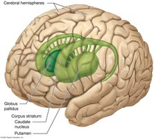
Basal nuclei are clusters of gray matter deep within the cerebral hemispheres. They play a crucial role in regulating voluntary movement and inhibiting involuntary movement.
Caudate Nucleus and Putamen (together, the corpus striatum)
Globus Pallidus: Inhibits inappropriate muscle contractions
Cerebral White Matter
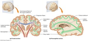
White matter in the cerebrum consists of three types of fibers:
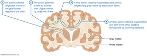
Commissural Fibers: Connect the two hemispheres (e.g., corpus callosum).
Projection Fibers: Connect the cortex with lower brain regions and the spinal cord.
Association Fibers: Connect regions within the same hemisphere.
Limbic System
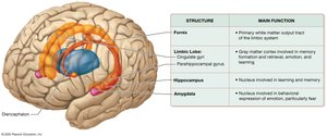
The limbic system is a functional group of structures involved in emotion, memory, and behavior. Key components include the limbic lobe, hippocampus, and amygdala.
Hippocampus: Memory formation
Amygdala: Emotional processing, especially fear
The Diencephalon
Major Components
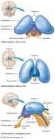
The diencephalon is located at the center of the brain and includes the thalamus, hypothalamus, epithalamus, and subthalamus.
Thalamus
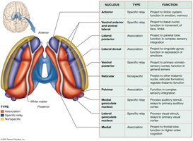
The thalamus acts as the main relay station for sensory and motor information to the cerebral cortex. It contains specific relay, association, and nonspecific nuclei, each with distinct functions in processing and integrating information.
Hypothalamus
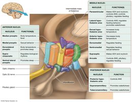
The hypothalamus regulates homeostatic functions such as hunger, thirst, body temperature, and the sleep/wake cycle. It is the "boss" of the autonomic nervous system and connects to the pituitary gland to control hormone secretion.
Epithalamus and Subthalamus
Epithalamus: Contains the pineal gland, which secretes melatonin for sleep regulation.
Subthalamus: Works with basal nuclei to control movement.
The Cerebellum
Structure and Function
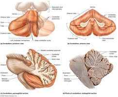
The cerebellum is located posteriorly and inferiorly in the brain, consisting of two hemispheres connected by the vermis. It coordinates movement, reduces motor error, and is essential for motor learning.
The Brainstem
Major Divisions and Functions
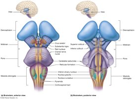
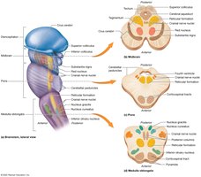
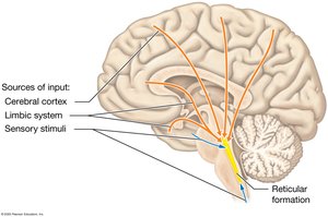
The brainstem connects the brain to the spinal cord and is divided into the midbrain, pons, and medulla oblongata. It controls basic life functions such as heart rate, breathing, and reflexes, and houses the reticular formation, which regulates arousal and consciousness.
Protection of the Brain
Cranial Meninges
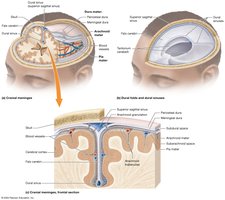
The brain is protected by three layers of connective tissue called meninges: dura mater (outermost), arachnoid mater (middle), and pia mater (innermost). These layers provide structural support and contain spaces filled with CSF for cushioning.
Ventricles and Cerebrospinal Fluid (CSF)
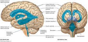
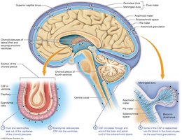
The brain contains four ventricles filled with CSF, which cushions the brain, removes waste, and maintains a stable environment. CSF is produced by the choroid plexuses and circulates through the ventricles and subarachnoid space.
Blood Brain Barrier
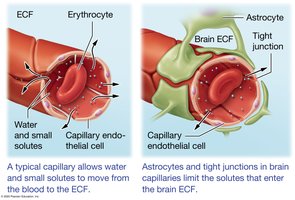
The blood brain barrier is formed by endothelial cells with tight junctions, astrocyte foot processes, and a basal lamina. It restricts the passage of harmful substances from the blood into the brain, while allowing essential molecules like oxygen and glucose to pass through.
The Spinal Cord
Structure and Function
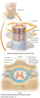
The spinal cord serves as a relay station for communication between the brain and the body and as a processing center for reflexes. It is protected by spinal meninges and contains both gray and white matter organized into horns and funiculi, respectively.
Module 1: Core Functions & Structural Map The Central Nervous System (CNS) processes sensory input, makes decisions, and coordinates physical movement. While the peripheral nervous system (PNS) handles basic sensory and motor pathways, integrative functions belong exclusively to the CNS . Licensed by Google Physical Metrics & Bounds Brain Weight : Generates an average mass of 1,250–1,450 g. Metabolic Demand : Consumes roughly 20% of total blood flow at rest. Spinal Cord Boundaries : Commences at the foramen magnum and terminates between the first and second lumbar vertebrae (\(L_1–L_2\)) .
Module 2: Gross Anatomy & Tissue Composition The brain houses four major subdivisions, each engineered for distinct functional tiers. ┌────────────────────────────────────────────────────────────────────────┐ │ FOUR DIVISION ARCHITECTURE │ ├───────────────┬──────────────────┬─────────────────┬───────────────────┤ │ CEREBRUM │ DIENCEPHALON │ CEREBELLUM │ BRAINSTEM │ ├───────────────┼──────────────────┼─────────────────┼───────────────────┤ │ Higher mental │ Homeostasis, │ Coordinates and │ Involuntary life │ │ functions, │ sensory relay, │ smooths motion, │ processes, basic │ │ logic, memory │ autonomic hub │ reduces error │ reflexes, arousal │ └───────────────┴──────────────────┴─────────────────┴───────────────────┘ Microscopic Organization White Matter : Made of myelinated axons that form long-distance communication tracts. Gray Matter : Made of neuron cell bodies, dendrites, and unmyelinated axons that form processing clusters. Brain Layout : Gray matter creates the superficial outer cortex, while white matter sits deep. Spinal Cord Layout : White matter sits on the surface (superficial), while gray matter forms an internal butterfly shape.
Module 3: Deep Cerebrum & Protective Barriers Internal Cerebral Systems Cerebral Cortex (Neocortex) : Formed by six distinct layers folded into gyri (ridges) and sulci/fissures (grooves). Basal Nuclei : Includes the caudate nucleus, putamen, and globus pallidus. These clusters inhibit involuntary movement and smooth out voluntary intention. Limbic System : Made up of the amygdala, hippocampus, limbic lobe, and fornix to govern emotion, learning, and memory. White Matter Fiber Types : Commissural Fibers : Bridges the left and right hemispheres (e.g., the corpus callosum ). Projection Fibers : Links the superficial cortex down to lower brain areas and the spinal cord. Association Fibers : Wire together different regions within the exact same hemisphere. The Triple Shield Protocol Cranial Meninges : Three protective membranes consisting of the outer dura mater , middle arachnoid mater , and inner pia mater . Cerebrospinal Fluid (CSF) : Produced by choroid plexuses inside four core brain ventricles to absorb impacts and flush cellular waste. Blood-Brain Barrier (BBB) : Created by endothelial cells with tight junctions to restrict toxic substances from leaking into neural tissue.
Module 4: Neuroembryology The entire adult CNS differentiates from an embryonic neural tube via primary and secondary vesicle stages: [Primary Vesicle] [Secondary Vesicle] [Adult CNS Structure] ├── Forebrain (Prosencephalon) ──┬── Telencephalon ──────────► Cerebrum │ └── Diencephalon ───────────► Thalamus & Hypothalamus │ ├── Midbrain (Mesencephalon) ────────────────────────────────► Midbrain (Brainstem) │ └── Hindbrain (Rhombencephalon) ─┬── Metencephalon ──────────► Pons & Cerebellum └── Myelencephalon ─────────► Medulla Oblongata
Summary Table: Major Brain Divisions and Functions
Brain Division | Function |
|---|---|
Cerebrum | Performs higher mental functions, interprets sensory stimuli, plans and initiates movement |
Diencephalon | Processes, integrates, and relays information; maintains homeostasis; regulates movement and biological rhythms |
Cerebellum | Monitors and coordinates movement |
Brainstem | Maintains homeostasis; controls certain reflexes; monitors movement; integrates and relays information |
