 Back
BackThe Central Nervous System: Structure, Function, and Integration
Study Guide - Smart Notes
 Tailored notes based on your materials, expanded with key definitions, examples, and context.
Overview of Central Nervous System (CNS) Functions
Functions of the Nervous System
The nervous system is responsible for detecting, integrating, and responding to internal and external stimuli. It is divided into sensory, integrative, and motor functions:
Sensory Functions: Detection of sensations within and outside the body.
Integrative Functions: Decision-making processes, primarily carried out by the CNS.
Motor Functions: Stimulation of muscle contractions or gland secretions.
Sensory and motor functions are performed by the peripheral nervous system (PNS), while integrative functions are exclusive to the CNS.
Basic Structure of the Brain and Spinal Cord
Major Divisions of the Brain
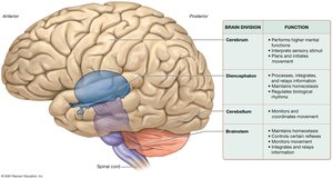
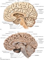
The brain is a soft, whitish-gray organ located in the cranial cavity, consisting of four main divisions:
Cerebrum: Responsible for higher mental functions such as learning, memory, personality, cognition, language, and conscience. It also plays a major role in sensation and movement.
Diencephalon: Processes, integrates, and relays information; maintains homeostasis, regulates movement, and biological rhythms.
Cerebellum: Coordinates and plans movement, especially complex activities.
Brainstem: Controls basic involuntary processes, mediates reflexes, monitors movement, and relays information.
White and Gray Matter
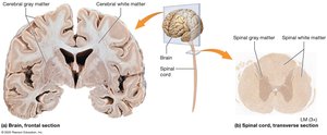
The CNS is organized into white and gray matter:
White Matter: Contains myelinated axons; forms tracts that transmit information.
Gray Matter: Composed of neuron cell bodies, dendrites, and unmyelinated axons; forms nuclei and the cerebral cortex.
In the brain, gray matter is superficial and scattered in deeper regions, while in the spinal cord, gray matter is internal and white matter is superficial.
Development of the CNS
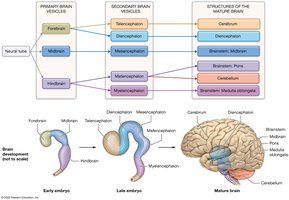
The CNS develops from a neural tube, which forms primary brain vesicles (forebrain, midbrain, hindbrain) that further differentiate into secondary vesicles and mature brain structures.
The Cerebrum
Structure and Lobes
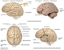
The cerebrum consists of two hemispheres, each divided into lobes:
Frontal Lobe: Involved in voluntary movement, planning, and higher cognitive functions.
Parietal Lobe: Processes sensory information related to touch, temperature, and proprioception.
Temporal Lobe: Involved in auditory processing and memory.
Occipital Lobe: Processes visual information.
Insula: Involved in taste and visceral sensation.
Cerebral Cortex and Functional Areas
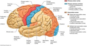
The neocortex is the most recent evolutionary part of the cerebral cortex, responsible for conscious processes. It contains:
Primary Motor Cortex: Plans and executes movement.
Primary Sensory Cortices: Process sensory input (e.g., somatosensory, visual, auditory).
Association Areas: Integrate multiple types of stimuli for higher-level processing.
Basal Nuclei
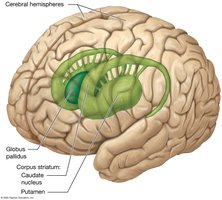
The basal nuclei are clusters of cell bodies involved in the regulation of movement and behavior. Major components include:
Caudate Nucleus
Putamen
Globus Pallidus
They inhibit involuntary movement and initiate voluntary movement.
Cerebral White Matter
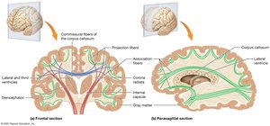
White matter in the cerebrum is organized into:
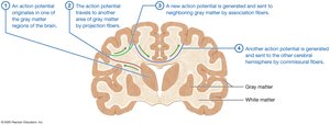
Commissural Fibers: Connect the two hemispheres (e.g., corpus callosum).
Projection Fibers: Connect the cortex with other brain regions and the spinal cord.
Association Fibers: Connect regions within the same hemisphere.
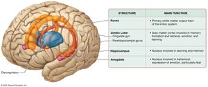
Limbic System
The limbic system is involved in memory, learning, emotion, and behavior. Key structures include:
Limbic Lobe
Hippocampus: Memory formation
Amygdala: Emotional processing, especially fear
The Diencephalon
Major Components
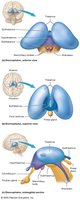
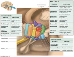
The diencephalon is centrally located and includes:
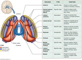
Thalamus: Main relay station for sensory information to the cortex.
Hypothalamus: Regulates homeostasis, endocrine functions, and the autonomic nervous system.
Epithalamus: Contains the pineal gland, which secretes melatonin.
Subthalamus: Involved in movement control.
The Cerebellum
Structure and Function
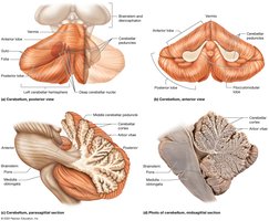
The cerebellum coordinates voluntary movement and maintains posture and balance. It consists of two hemispheres connected by the vermis and is divided into lobes. The arbor vitae is the white matter of the cerebellum.
The Brainstem
Major Divisions and Functions
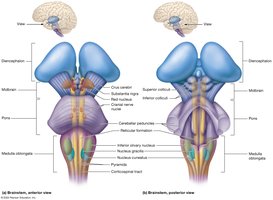
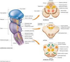
The brainstem connects the brain to the spinal cord and is divided into:
Midbrain: Involved in movement, reflexes, and processing visual/auditory information.
Pons: Relays information and regulates breathing and sleep.
Medulla Oblongata: Controls heart rate, breathing, and other autonomic functions.
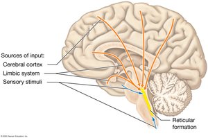
Reticular Formation
The reticular formation is a network of nuclei involved in arousal, consciousness, and regulation of vital functions such as heart rate and respiration.
Protection of the Brain
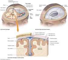
Cranial Meninges
The brain is protected by three layers of connective tissue called meninges:
Dura Mater: Outermost, tough layer.
Arachnoid Mater: Middle, web-like layer.
Pia Mater: Innermost, delicate layer adhering to the brain surface.
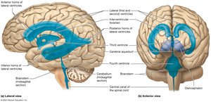
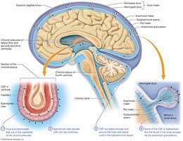
Ventricles and Cerebrospinal Fluid (CSF)
The brain contains interconnected cavities called ventricles, filled with CSF, which cushions the brain, removes waste, and maintains homeostasis.
Blood Brain Barrier
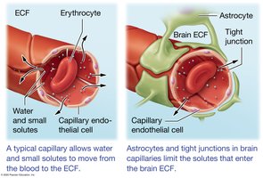
The blood brain barrier is formed by endothelial cells with tight junctions, astrocyte foot processes, and a basal lamina. It restricts the passage of substances from the blood to the brain, protecting neural tissue from toxins and pathogens.
The Spinal Cord
Structure and Protection
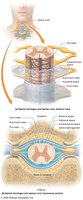
The spinal cord is a long, tubular structure encased within the vertebral column. It is protected by spinal meninges (dura, arachnoid, pia mater) and spaces (epidural, subdural, subarachnoid).
External and Internal Anatomy
The spinal cord has external features such as the conus medullaris, filum terminale, and cauda equina. Internally, it contains butterfly-shaped gray matter surrounded by white matter tracts.
Sensation and Perception
General Somatic Senses
General somatic senses include touch, stretch, joint position, pain, and temperature. Sensory pathways include the posterior columns/medial lemniscal system and the anterolateral system (spinothalamic tracts).
Movement and Motor Pathways
Voluntary Movement
Voluntary movement is planned and coordinated by the motor areas of the cerebral cortex, basal nuclei, cerebellum, and spinal cord. Motor pathways include the corticospinal and corticonuclear tracts.
Summary Table: Major Brain Divisions and Functions
Brain Division | Function |
|---|---|
Cerebrum | Performs higher mental functions, interprets sensory stimuli, plans and initiates movement |
Diencephalon | Processes, integrates, and relays information; maintains homeostasis; regulates movement and biological rhythms |
Cerebellum | Monitors and coordinates movement |
Brainstem | Maintains homeostasis, controls certain reflexes, monitors movement, integrates and relays information |
