 Back
BackThe Central Nervous System: Structure, Function, and Integration
Study Guide - Smart Notes
 Tailored notes based on your materials, expanded with key definitions, examples, and context.
Overview of the Central Nervous System (CNS)
Definition and General Functions
The central nervous system (CNS) consists of the brain and spinal cord. It is responsible for integrating sensory information, coordinating movement, maintaining homeostasis, and supporting higher mental functions such as learning, memory, and language.
Motor functions: Initiate muscle contraction or gland secretion (primarily via the peripheral nervous system, PNS).
Sensory functions: Detect sensations inside and outside the body (also primarily via the PNS).
Integrative functions: Decision-making processes, including interpretation of sensory input, planning movement, maintaining homeostasis, and higher mental functions (exclusive to the CNS).
Basic Structure of the Brain and Spinal Cord
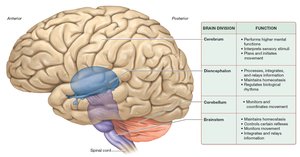
The brain is a soft, whitish-gray organ located in the cranial cavity, weighing 1250–1450 grams. It is composed mainly of nervous tissue, with some epithelial and connective tissues. The brain receives about 20% of the body's blood flow at rest, reflecting its high demand for oxygen and nutrients. The brain contains four main divisions:
Cerebrum
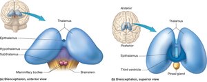
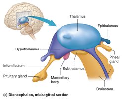
Diencephalon
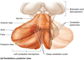
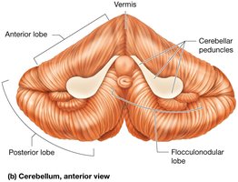
Cerebellum
Brainstem
White and Gray Matter
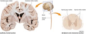
Both the brain and spinal cord contain white matter (myelinated axons) and gray matter (neuron cell bodies, dendrites, and unmyelinated axons). In the brain, gray matter is superficial (cerebral cortex), while in the spinal cord, gray matter is central and white matter is superficial.
The Brain
Cerebrum
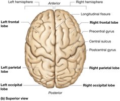
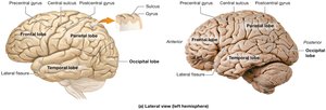
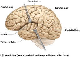
The cerebrum is the largest part of the brain, responsible for higher mental functions such as learning, memory, personality, cognition, language, and conscience. It is divided into left and right hemispheres, each with five lobes:
Frontal lobe: Planning and executing movement, behavior, conscience, personality
Parietal lobe: Processing and integrating sensory information, attention
Temporal lobe: Hearing, language, memory, emotions
Occipital lobe: Vision
Insula: Taste and visceral functions
Surface Features
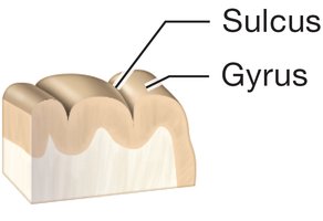
Gyri: Elevated ridges
Sulci: Shallow grooves
Fissures: Deep grooves (e.g., longitudinal fissure separates hemispheres)
Cerebral Cortex
The cerebral cortex is the outer layer of gray matter, mainly composed of interneurons. It is divided into:
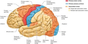
Primary motor cortex: Plans and executes movement
Primary sensory cortices: Receive and process sensory input
Association areas: Integrate information (unimodal for one type, multimodal for multiple types)
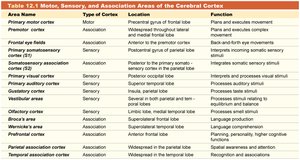
Area Name | Type of Cortex | Location | Function |
|---|---|---|---|
Primary motor cortex | Motor | Precentral gyrus of frontal lobe | Plans and executes movement |
Frontal eye fields | Association | Anterior to premotor cortex | Back-and-forth eye movements |
Primary somatosensory cortex (S1) | Sensory | Postcentral gyrus of parietal lobe | Processes somatic sensory stimuli |
Somatosensory association cortex (S2) | Association | Posterior to S1 | Integrates somatic sensory stimuli |
Primary visual cortex | Sensory | Occipital lobe | Processes visual stimuli |
Visual association area | Association | Occipital lobe | Processes and integrates visual stimuli |
Primary auditory cortex | Sensory | Superior temporal lobe | Processes auditory stimuli |
Auditory association area | Association | Superior temporal lobe | Processes and integrates auditory stimuli |
Gustatory cortex | Sensory | Insula and parietal lobe | Processes taste stimuli |
Vestibular areas | Sensory | Parietal and temporal lobes | Processes equilibrium stimuli |
Olfactory cortex | Sensory | Limbic and medial temporal lobes | Processes smell stimuli |
Prefrontal cortex | Association | Widespread in frontal lobe | Planning, personality, higher cognitive functions |
Parietal association cortex | Association | Widespread in parietal lobe | Integration, spatial awareness, attention |
Temporal association cortex | Association | Widespread in temporal lobe | Recognition and association |
Basal Nuclei
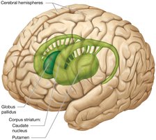
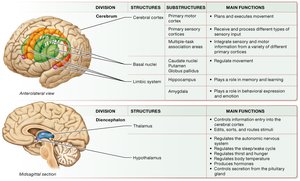
The basal nuclei are clusters of neuron cell bodies deep within each hemisphere, involved in movement regulation. Major components include the caudate nucleus, putamen, and globus pallidus.
Cerebral White Matter
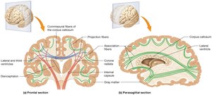
Commissural fibers: Connect right and left hemispheres (e.g., corpus callosum)
Projection fibers: Connect cortex with other brain regions and spinal cord
Association fibers: Connect gyri within a single hemisphere
Limbic System
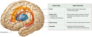
The limbic system is involved in memory, learning, emotion, and behavior. It includes the limbic lobe, hippocampus, amygdala, and associated pathways.
Diencephalon
Major Components
Thalamus: Main entry route for sensory data to the cortex; relay and processing center
Hypothalamus: Regulates autonomic functions, endocrine activity, body temperature, and homeostasis
Epithalamus: Contains pineal gland, which secretes melatonin
Subthalamus: Involved in movement control with basal nuclei
Cerebellum
Structure and Function
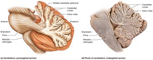
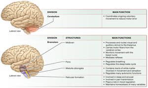
The cerebellum coordinates movement and maintains balance. It is divided into two hemispheres and three lobes (anterior, posterior, flocculonodular). The cerebellar cortex is highly folded, and the inner white matter forms the arbor vitae.
Brainstem
Structure and Function
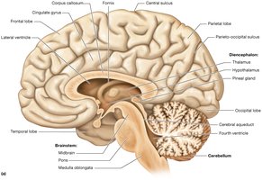
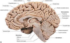
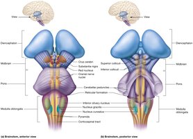
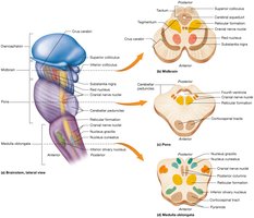
The brainstem connects the brain to the spinal cord and is essential for basic life functions. It consists of the midbrain, pons, and medulla oblongata. The brainstem contains nuclei for cranial nerves, the reticular formation, and tracts for sensory and motor pathways.
Reticular Formation
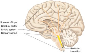
The reticular formation is a network of nuclei in the brainstem involved in regulating sleep, consciousness, pain, and homeostasis.
Protection of the Brain
Cranial Meninges
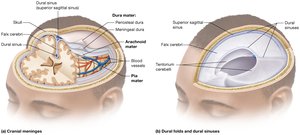
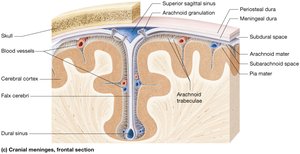
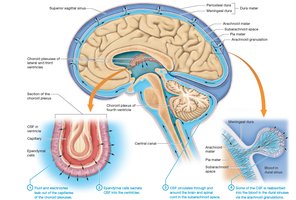
The brain is protected by three layers of meninges: dura mater, arachnoid mater, and pia mater. These layers provide structural support and contain spaces for cerebrospinal fluid (CSF) and blood vessels.
Ventricles and Cerebrospinal Fluid (CSF)
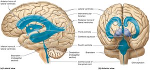
The brain contains four ventricles filled with CSF, which cushions the brain, removes waste, and maintains a stable environment. CSF is produced by the choroid plexuses and circulates through the ventricles and subarachnoid space.
Blood-Brain Barrier
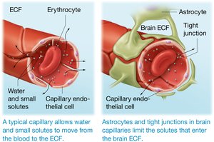
The blood-brain barrier is formed by endothelial cells with tight junctions, astrocytes, and basal laminae. It restricts the passage of harmful substances from the blood into the brain while allowing essential nutrients to pass.
The Spinal Cord
Structure and Protection
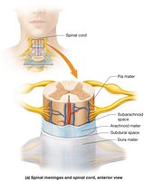
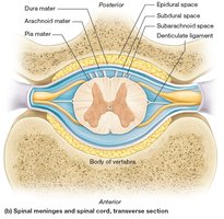
The spinal cord is a long, tubular structure that relays information between the brain and body and processes some reflexes. It is protected by spinal meninges (dura mater, arachnoid mater, pia mater) and spaces (epidural, subdural, subarachnoid).
Summary Table: Major Brain Structures and Their Functions
Division | Structures | Main Functions |
|---|---|---|
Cerebrum | Primary motor cortex, sensory cortices, association areas, basal nuclei, limbic system | Plans and executes movement, processes sensory input, integrates information, memory, learning, emotion |
Diencephalon | Thalamus, hypothalamus | Relays sensory input, regulates homeostasis, controls endocrine functions |
Cerebellum | Cerebellar cortex, deep nuclei | Coordinates movement, maintains balance |
Brainstem | Midbrain, pons, medulla oblongata, reticular formation | Controls vital functions, relays information, regulates consciousness |
Additional info: This summary covers the structure and function of the central nervous system, including the brain and spinal cord, their protective features, and the integration of sensory and motor functions. It is suitable for college-level Anatomy & Physiology students preparing for exams on the CNS.
