 Back
BackThe Central Nervous System: Structure, Function, and Integration
Study Guide - Smart Notes
 Tailored notes based on your materials, expanded with key definitions, examples, and context.
Overview of Central Nervous System (CNS) Functions
Major Functions of the Nervous System
The nervous system is responsible for detecting, integrating, and responding to internal and external stimuli. It is divided into sensory, integrative, and motor functions:
Sensory Functions: Detection of sensations within and outside the body.
Integrative Functions: Decision-making processes, primarily carried out by the CNS.
Motor Functions: Stimulation of muscle contractions or gland secretions.
Sensory and motor functions are performed by the peripheral nervous system (PNS), while integrative functions are exclusive to the CNS.
Basic Structure of the Brain and Spinal Cord
Major Divisions of the Brain
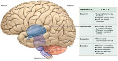
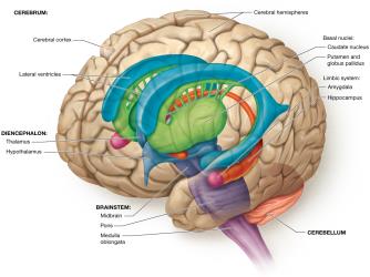
The brain is a soft, whitish-gray organ located in the cranial cavity, composed mainly of nervous tissue, with some connective and modified epithelial tissue. It contains internal cavities called ventricles, filled with cerebrospinal fluid (CSF), and is divided into four main regions:
Cerebrum: Responsible for higher mental functions, sensation, and movement.
Diencephalon: Processes, integrates, and relays information; maintains homeostasis and regulates biological rhythms.
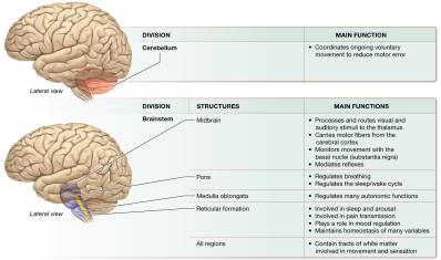
Cerebellum: Coordinates movement and motor learning.
Brainstem: Maintains homeostasis, controls reflexes, and relays information between the brain and spinal cord.
Spinal Cord
The spinal cord is a long, tubular organ encased within the vertebral cavity, beginning at the foramen magnum and ending between the first and second lumbar vertebrae. It serves as a relay and processing station for neural signals and contains a central canal filled with CSF.
White and Gray Matter in the CNS
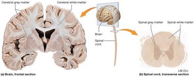
The CNS is organized into white and gray matter:
White Matter: Contains myelinated axons; in the brain, it is deep to the cortex, while in the spinal cord, it is superficial.
Gray Matter: Composed of neuron cell bodies, dendrites, and unmyelinated axons; forms the cortex and nuclei in the brain, and is internal in the spinal cord.
Development of the CNS
Neural Tube and Brain Vesicles
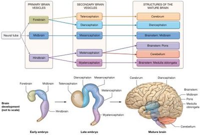
The CNS develops from a hollow neural tube. By the fourth week of development, the cranial end forms three primary brain vesicles (forebrain, midbrain, hindbrain), which further differentiate into five secondary brain vesicles by the fifth week. These vesicles give rise to the mature structures of the brain.
The Cerebrum
Structure and Lobes
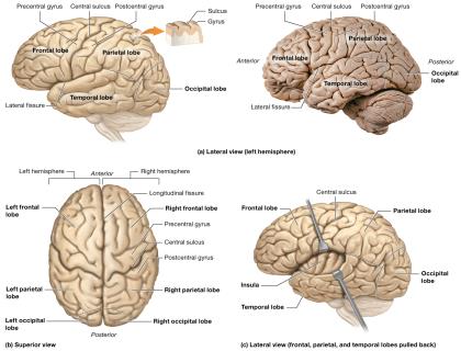
The cerebrum consists of two hemispheres, each divided into lobes:
Frontal Lobe: Involved in voluntary movement, planning, and higher cognitive functions.
Parietal Lobe: Processes sensory information related to touch, temperature, and proprioception.
Temporal Lobe: Involved in auditory processing and memory.
Occipital Lobe: Responsible for visual processing.
Insula: Located deep within the lateral fissure; involved in taste and visceral sensation.
Cerebral Cortex and Functional Areas
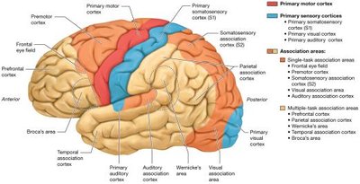
The cerebral cortex is the outer layer of gray matter, responsible for conscious processes. It contains several functional areas:
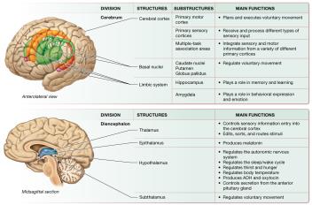
Primary Motor Cortex: Plans and executes movement.
Primary Sensory Cortices: Process sensory input (e.g., somatosensory, visual, auditory).
Association Areas: Integrate different types of information for complex processing.
Basal Nuclei
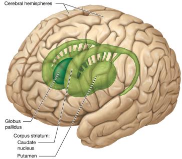
The basal nuclei are clusters of gray matter deep within the cerebral hemispheres, including the caudate nucleus, putamen, and globus pallidus. They regulate voluntary movement and inhibit involuntary movement.
Cerebral White Matter
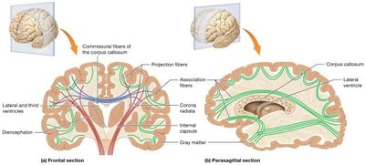
White matter in the cerebrum consists of:
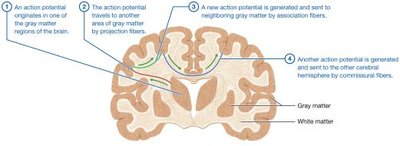
Commissural Fibers: Connect the two hemispheres (e.g., corpus callosum).
Projection Fibers: Connect the cortex with lower brain regions and the spinal cord.
Association Fibers: Connect regions within the same hemisphere.
Limbic System
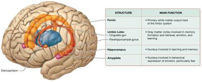
The limbic system is involved in memory, learning, emotion, and behavior. Major components include the limbic lobe, hippocampus, and amygdala.
The Diencephalon
Major Components
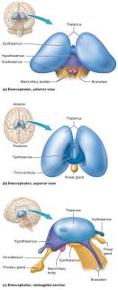
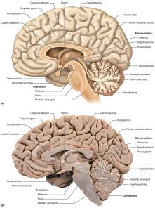
The diencephalon is located at the center of the brain and includes:
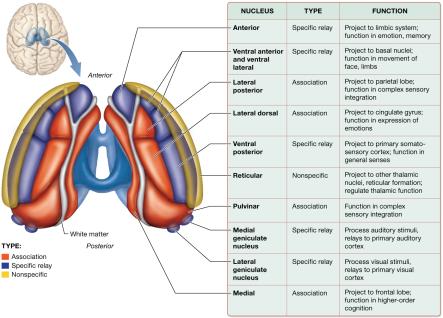
Thalamus: Main relay station for sensory information to the cortex.
Epithalamus: Contains the pineal gland, which secretes melatonin.
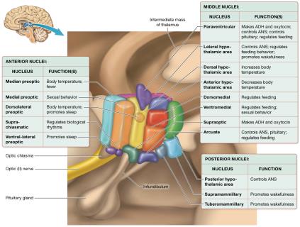
Hypothalamus: Regulates homeostasis, the autonomic nervous system, and the endocrine system.
Subthalamus: Involved in movement regulation.
The Cerebellum
Structure and Function
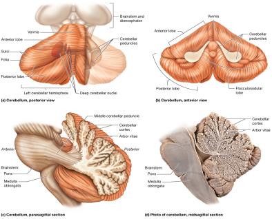
The cerebellum is located posterior and inferior to the cerebrum. It coordinates voluntary movement, reduces motor error, and is involved in motor learning. It consists of two hemispheres connected by the vermis and is divided into three lobes.
The Brainstem
Major Divisions and Functions
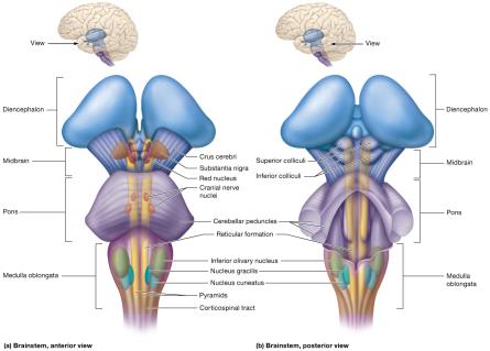
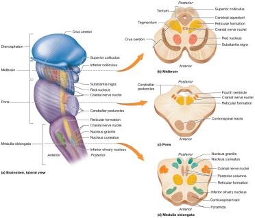
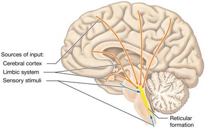
The brainstem connects the brain to the spinal cord and is divided into the midbrain, pons, and medulla oblongata. It controls basic life functions such as heart rate, breathing, and reflexes, and contains the reticular formation, which regulates arousal and consciousness.
Integration of Brain Structures and Functions
Major brain structures work together to regulate voluntary movement, sensation, cognition, and homeostasis. The cerebrum, diencephalon, cerebellum, and brainstem each contribute specialized functions, but are highly interconnected.
Sleep and Wakefulness
Circadian Rhythms and the Biological Clock
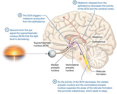
Sleep is regulated by the hypothalamic suprachiasmatic nucleus (SCN), which acts as the biological clock. The SCN receives input about light levels and triggers melatonin production from the epithalamus, which decreases cortical activity and promotes sleep.
Brain Waves and Stages of Sleep
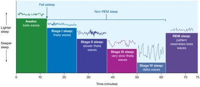
Sleep consists of several stages, each characterized by distinct brain wave patterns measured by EEG:
Beta Waves: Awake, alert state.
Theta Waves: Light sleep (Stages I–III).
Delta Waves: Deep sleep (Stage IV).
REM Sleep: Rapid eye movement, dreaming, muscle paralysis.
Additional info: The CNS is essential for integrating sensory input, coordinating voluntary and involuntary responses, and maintaining homeostasis. Its complex structure allows for specialization of function, but also requires extensive communication between regions for normal function.
