 Back
BackThe Central Nervous System: Structure, Function, and Organization
Study Guide - Smart Notes
 Tailored notes based on your materials, expanded with key definitions, examples, and context.
Chapter 12 – The Central Nervous System
Overview of the Central Nervous System (CNS)
The central nervous system (CNS) is composed of the brain and spinal cord, serving as the main control center for the body. It integrates sensory information, coordinates voluntary and involuntary responses, and is responsible for higher mental functions such as thought, memory, and emotion.
Brain Regions and Organization
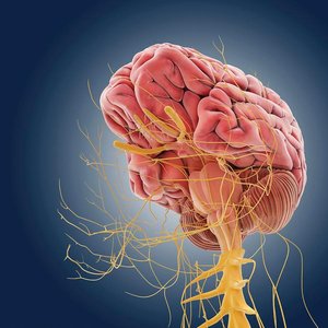
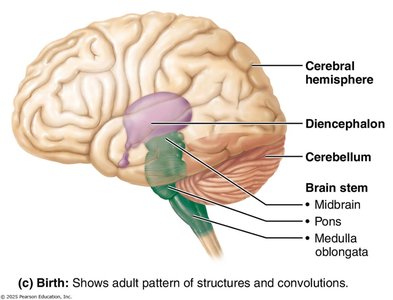
The brain is divided into several major regions, each with specialized functions. These include the cerebrum, diencephalon, cerebellum, and brain stem. The brain stem is further subdivided into the midbrain, pons, and medulla oblongata. The spinal cord extends from the brain stem and is protected within the vertebral canal.
Higher nerve centers: Cerebrum, diencephalon, cerebellum, and part of the brain stem
Lower nerve centers: Brain stem (in part) and spinal cord
Gray matter: Contains neuron cell bodies; forms the cortex in the cerebrum and cerebellum
White matter: Composed of myelinated fibers; found externally in the spinal cord and internally in the brain
Brain Ventricles and Cerebrospinal Fluid (CSF)
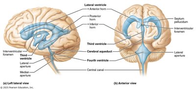
The brain contains a network of interconnected cavities called ventricles, which are filled with cerebrospinal fluid (CSF). CSF cushions the brain, provides nutrients, and removes waste. The ventricles are lined by ependymal cells that help circulate CSF.
Lateral ventricles: C-shaped, located in the cerebral hemispheres, separated by the septum pellucidum
Third ventricle: Located in the diencephalon
Fourth ventricle: Located between the brain stem and cerebellum
Cerebral Hemispheres
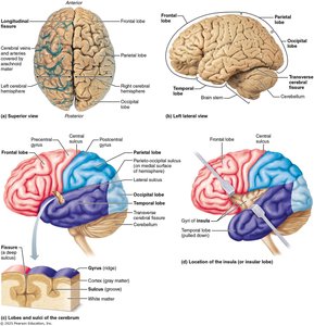
The cerebrum is divided into two hemispheres, each containing five lobes: frontal, parietal, temporal, occipital, and insula. The hemispheres are separated by the longitudinal fissure and connected internally by the corpus callosum. Surface markings such as sulci and gyri define the lobes and functional areas.
Cerebral cortex: Outer layer of gray matter responsible for conscious thought and voluntary actions
White matter: Internal axonal tracts
Basal nuclei: Deep islands of gray matter involved in movement and cognition
Cerebral Cortex: Structure and Function
The cerebral cortex is the site of the conscious mind, responsible for awareness, sensory perception, voluntary motor activities, communication, memory, and understanding. It is organized into functional areas, each with specialized roles.
Contralateral control: Each hemisphere controls the opposite side of the body
Prefrontal cortex: Involved in intellect, complex learning, recall, and personality
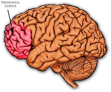
Motor Areas of the Cerebral Cortex
Motor areas are responsible for voluntary movement and are located in the frontal lobe.
Primary motor cortex: Located in the precentral gyrus; controls precise, voluntary movements
Premotor cortex: Plans and sequences complex movements
Broca’s area: Controls muscles involved in speech production
Frontal eye field: Controls voluntary eye movements
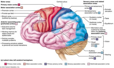
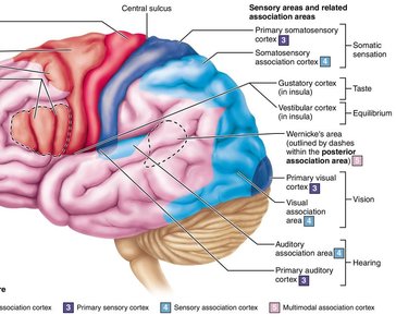
Sensory Areas of the Cerebral Cortex
Sensory areas receive and interpret sensory information from the body and environment.
Primary somatosensory cortex: Receives information from skin and proprioceptors
Visual cortex: Processes visual information
Auditory cortex: Processes sound
Olfactory cortex: Perception of odors
Gustatory cortex: Perception of taste
Visceral sensory area: Perception of visceral sensations (e.g., full bladder)
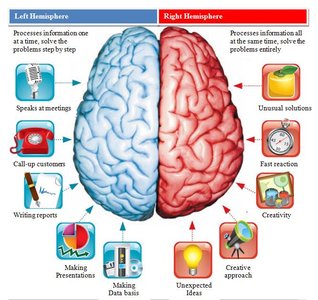
Lateralization of Cortical Function
Although the cerebral hemispheres are structurally similar, they exhibit functional specialization, known as lateralization.
Left hemisphere: Language, math, logic
Right hemisphere: Visual-spatial skills, intuition, emotion, artistic and musical abilities
Hemispheres communicate via fiber tracts such as the corpus callosum
Cerebral White Matter
Cerebral white matter consists of myelinated fibers that facilitate communication within the brain and between the brain and spinal cord.
Association fibers: Connect different parts of the same hemisphere
Commissural fibers: Connect corresponding gray areas of the two hemispheres (e.g., corpus callosum)
Projection fibers: Connect the cortex with lower brain regions and spinal cord
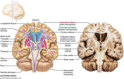
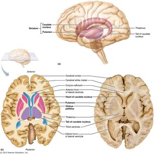
Basal Nuclei (Basal Ganglia)
The basal nuclei are deep gray matter structures involved in the regulation of movement, cognition, and emotion. They filter out inappropriate responses and send the best response to the cortex. Disorders such as Parkinson’s and Huntington’s disease are associated with basal nuclei dysfunction.
Components: Caudate nucleus, putamen, globus pallidus
Functions: Movement regulation, cognitive and emotional roles
Diencephalon
The diencephalon is composed of the thalamus, hypothalamus, and epithalamus, surrounding the third ventricle. It acts as a relay and processing center for sensory and autonomic information.
Thalamus: Relay station for sensory and motor signals to the cerebral cortex
Hypothalamus: Main visceral control center; regulates homeostasis, autonomic functions, emotions, body temperature, hunger, thirst, sleep-wake cycles, and endocrine system
Epithalamus: Contains the pineal gland, which secretes melatonin to regulate sleep-wake cycles
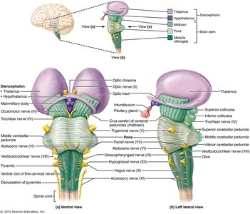
Brain Stem
The brain stem consists of the midbrain, pons, and medulla oblongata. It controls automatic survival functions and connects higher and lower nervous system centers.
Midbrain: Contains visual and auditory relay centers, substantia nigra (dopamine production), and red nucleus (motor coordination)
Pons: Relays information between the cerebrum and cerebellum; involved in breathing regulation
Medulla oblongata: Autonomic reflex center; regulates cardiovascular, respiratory, and other vital functions
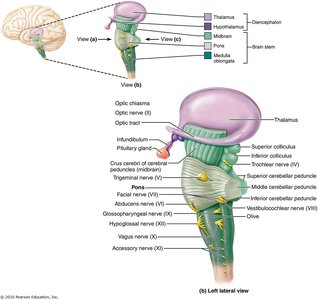
Reticular Formation
The reticular formation is a network of neurons extending through the brain stem. It regulates arousal, attention, and consciousness via the reticular activating system (RAS). It also filters sensory input and controls skeletal muscle and visceral motor functions.
Cerebellum
The cerebellum is located dorsal to the pons and medulla and is responsible for coordinating movement, balance, and posture. It receives input from the cortex, brain stem, and sensory receptors to fine-tune motor activity.
Structure: Two hemispheres, each with three lobes (anterior, posterior, flocculonodular)
Arbor vitae: Tree-like arrangement of white matter
Sleep and Sleep/Wake Cycles
Sleep is a state of partial unconsciousness with two main types: non-rapid eye movement (NREM) and rapid eye movement (REM) sleep. Sleep cycles involve transitions through NREM stages and REM, with physiological changes such as decreased heart rate and blood pressure during NREM and increased brain activity during REM.
Protection of the Brain
The brain is protected by the skull, meninges (dura mater, arachnoid mater, pia mater), cerebrospinal fluid, and the blood-brain barrier. The meninges cover and protect the CNS, contain CSF, and form partitions within the skull. The blood-brain barrier restricts passage of harmful substances from the blood to the brain.
Homeostatic Imbalances of the CNS
Disorders of the CNS include traumatic injuries (concussion, contusion, hemorrhage), cerebrovascular accidents (stroke, ischemia, hemiplegia), and degenerative diseases (Alzheimer’s, Parkinson’s, Huntington’s). These conditions can result in loss of function, memory, movement, or even death.
Spinal Cord: Structure and Function
The spinal cord extends from the foramen magnum to the L1/L2 vertebra and serves as a communication pathway between the brain and body. It contains reflex centers and is protected by vertebrae, meninges, and CSF.
31 pairs of spinal nerves: Emerge from the spinal cord to serve the body
Gray matter: Arranged in dorsal (sensory) and ventral (motor) horns
White matter: Contains ascending (sensory), descending (motor), and transverse (commissural) tracts
Spinal Cord Trauma
Injuries to the spinal cord can result in sensory and motor deficits. Complete transection leads to loss of function below the injury site. Paraplegia and quadriplegia are determined by the level of injury. Regeneration is limited due to inhibitory factors in the CNS.
Summary Table: Major Brain Regions and Functions
Region | Main Functions |
|---|---|
Cerebrum | Conscious thought, voluntary movement, sensory perception, memory, language |
Diencephalon | Relay of sensory/motor signals, homeostasis, endocrine regulation |
Cerebellum | Coordination of movement, balance, posture |
Brain Stem | Autonomic functions, relay between brain and spinal cord |
Spinal Cord | Reflexes, communication between brain and body |
Additional info: The above notes integrate and expand upon the provided lecture slides and images, ensuring a comprehensive, exam-ready summary of the central nervous system for Anatomy & Physiology students.
