Back
BackThe Central Nervous System: Structure, Function, and Protection
Study Guide - Smart Notes
 Tailored notes based on your materials, expanded with key definitions, examples, and context.
The Central Nervous System (CNS)
Overview of CNS Functions
The central nervous system (CNS) is responsible for integrating sensory information, making decisions, and coordinating motor output. It consists of the brain and spinal cord, which work together to maintain homeostasis and enable complex behaviors.
Sensory Functions: Detection of sensations inside and outside the body.
Integrative Functions: Decision-making processes, performed exclusively by the CNS.
Motor Functions: Stimulation of muscle contractions or gland secretions.
Basic Structure of the Brain and Spinal Cord
The brain is a soft, whitish-gray organ located in the cranial cavity, divided into four main regions. The spinal cord is a long, tubular structure encased within the vertebral cavity, connecting the brain to the peripheral nervous system.
Brain: Contains internal cavities called ventricles filled with cerebrospinal fluid (CSF). It receives about 20% of the body's blood flow at rest.
Spinal Cord: Begins at the foramen magnum and ends between the first and second lumbar vertebrae. The central canal is filled with CSF and is continuous with the brain's ventricles.
White Matter and Gray Matter
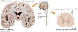
White matter consists of myelinated axons, while gray matter is composed of neuron cell bodies, dendrites, and unmyelinated axons. Their arrangement differs between the brain and spinal cord.
Brain: Gray matter forms the outer cortex and is scattered in deeper regions; white matter tracts connect different brain areas.
Spinal Cord: Gray matter is internal, surrounded by superficial white matter tracts.
Development of the CNS
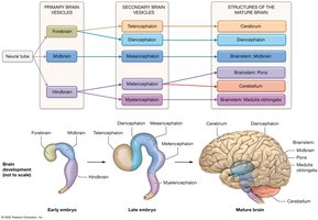
The CNS develops from a neural tube, which forms primary brain vesicles that later differentiate into secondary vesicles and mature brain structures.
Primary Brain Vesicles: Forebrain, Midbrain, Hindbrain.
Secondary Brain Vesicles: Telencephalon, Diencephalon, Mesencephalon, Metencephalon, Myelencephalon.
Mature Structures: Cerebral hemispheres, diencephalon, midbrain, pons, cerebellum, medulla oblongata.
The Cerebrum
Structure and Lobes
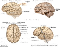
The cerebrum is the largest part of the brain, divided into right and left hemispheres. Its surface is marked by gyri (ridges), sulci (shallow grooves), and fissures (deep grooves). The hemispheres are separated by the longitudinal fissure.
Frontal Lobe: Anterior region; involved in voluntary movement, planning, and higher cognitive functions.
Parietal Lobe: Posterior to the frontal lobe; processes sensory information.
Temporal Lobe: Lateral surface; involved in auditory processing and memory.
Occipital Lobe: Posterior region; processes visual information.
Insula: Deep within the lateral fissure; involved in taste and visceral sensation.
Cerebral Cortex
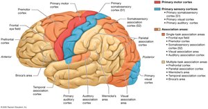
The cerebral cortex is the outer layer of gray matter, responsible for conscious processes. It contains specialized areas for motor, sensory, and association functions.
Primary Motor Cortex: Plans and executes voluntary movements.
Primary Sensory Cortices: Process sensory input (e.g., somatosensory, visual, auditory).
Association Areas: Integrate and interpret sensory and motor information.
Basal Nuclei
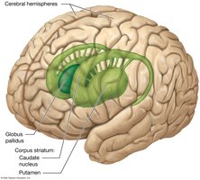
Basal nuclei are clusters of gray matter deep within the cerebral hemispheres. They regulate voluntary movement and inhibit involuntary movement.
Caudate Nucleus: C-shaped structure involved in movement regulation.
Putamen: Works with the caudate nucleus as part of the corpus striatum.
Globus Pallidus: Inhibits inappropriate muscle contractions.
Cerebral White Matter
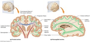
White matter in the cerebrum consists of myelinated axons organized into three types of fibers:
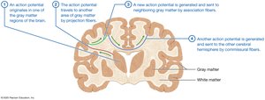
Commissural Fibers: Connect the two hemispheres (e.g., corpus callosum).
Projection Fibers: Connect the cortex with lower brain regions and the spinal cord.
Association Fibers: Connect regions within the same hemisphere.
Limbic System
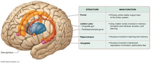
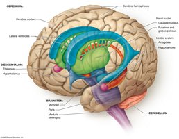
The limbic system is a functional group of structures involved in emotion, memory, and behavior. It includes the limbic lobe, hippocampus, amygdala, and connecting pathways.
Hippocampus: Essential for memory formation.
Amygdala: Involved in emotional responses, especially fear.
The Diencephalon
Structure and Components
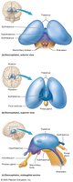
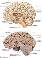
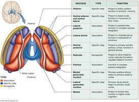
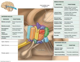
The diencephalon is located at the center of the brain and consists of the thalamus, hypothalamus, epithalamus, and subthalamus.
Thalamus: Main relay station for sensory information to the cortex.
Hypothalamus: Regulates homeostasis, autonomic functions, and endocrine activity.
Epithalamus: Contains the pineal gland, which secretes melatonin.
Subthalamus: Involved in movement control.
The Cerebellum
Structure and Function
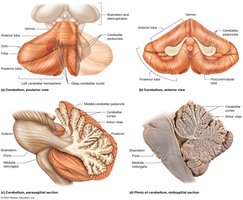
The cerebellum is located posterior and inferior to the cerebrum. It coordinates movement and helps maintain balance and posture.
Cerebellar Hemispheres: Connected by the vermis.
Folia: Surface ridges separated by sulci.
Arbor Vitae: White matter tracts within the cerebellum.
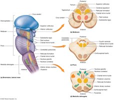
The Brainstem
Structure and Divisions
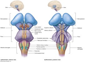
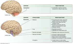
The brainstem connects the brain to the spinal cord and controls vital functions such as heart rate and breathing. It consists of the midbrain, pons, and medulla oblongata, and contains the reticular formation.
Midbrain: Contains nuclei involved in movement and reflexes.
Pons: Relays information and regulates breathing and sleep.
Medulla Oblongata: Controls autonomic functions and relays sensory information.
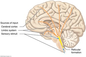
Reticular Formation: Regulates arousal, consciousness, and basic functions.
Major Brain Structures and Their Functions
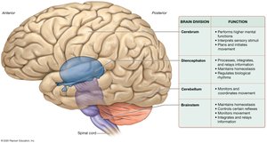
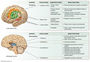
The brain's major divisions and their primary functions can be summarized as follows:
Division | Main Function |
|---|---|
Cerebrum | Performs higher mental functions, interprets sensory stimuli, plans and initiates movement |
Diencephalon | Processes, integrates, and relays information; maintains homeostasis; regulates movement and biological rhythms |
Cerebellum | Monitors and coordinates movement |
Brainstem | Maintains homeostasis; controls certain reflexes; monitors movement; integrates and relays information |
Protection of the Brain
Cranial Meninges
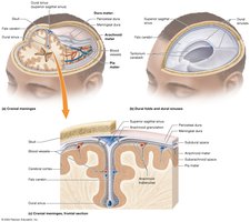
The brain is protected by three connective tissue membranes called meninges:
Dura Mater: Outermost, tough layer; forms dural sinuses for venous drainage.
Arachnoid Mater: Middle, web-like layer; contains the subarachnoid space filled with CSF.
Pia Mater: Innermost, thin layer; adheres to the brain surface.
The Ventricles and Cerebrospinal Fluid (CSF)
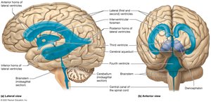
The brain contains four ventricles filled with CSF, which cushions the brain, removes waste, and maintains a stable environment.
Lateral Ventricles: Located in each cerebral hemisphere.
Third Ventricle: Located in the diencephalon.
Fourth Ventricle: Located between the pons and cerebellum.
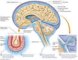
CSF Formation: Produced by choroid plexuses, circulates through ventricles and subarachnoid space, reabsorbed by arachnoid granulations.
The Blood Brain Barrier
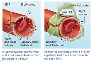
The blood brain barrier is formed by endothelial cells with tight junctions, supported by astrocytes. It restricts the passage of substances from the blood to the brain, protecting neural tissue from toxins and pathogens while allowing essential nutrients to pass.
Freely Permeable: Water, oxygen, carbon dioxide, lipid-soluble substances.
Selective Transport: Glucose, amino acids, ions via channels or carriers.
Restricted: Large, polar molecules and many drugs.