 Back
The Central Nervous System: Structure, Function, and Organization
Study Guide - Smart Notes
 Tailored notes based on your materials, expanded with key definitions, examples, and context.
Overview of the Central Nervous System (CNS)
Functions of the Nervous System
The nervous system is responsible for detecting sensations, integrating information, and initiating motor responses. It is divided into the central nervous system (CNS) and peripheral nervous system (PNS). The CNS, consisting of the brain and spinal cord, is the primary site for integrative functions, while the PNS handles sensory and motor functions.
Sensory Functions: Detection of internal and external stimuli.
Integrative Functions: Processing and interpretation of sensory input, decision-making.
Motor Functions: Initiation of muscle contraction or gland secretion.
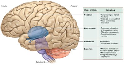
Basic Structure of the Brain and Spinal Cord
Gross Anatomy
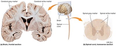
The brain is a soft, whitish-gray organ housed in the cranial cavity, composed mainly of nervous tissue, with some connective and epithelial tissue. It contains internal cavities called ventricles, filled with cerebrospinal fluid (CSF). The spinal cord is a long, thin structure extending from the brainstem through the vertebral canal.
White Matter: Myelinated axons; forms tracts for communication.
Gray Matter: Neuron cell bodies, dendrites, and unmyelinated axons; forms nuclei and cortex.
Development of the Brain
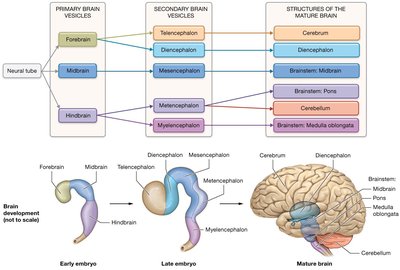
The brain develops from the neural tube, forming primary and secondary brain vesicles, which give rise to the mature brain structures: cerebrum, diencephalon, brainstem, and cerebellum.
The Cerebrum
Lobes and Surface Features
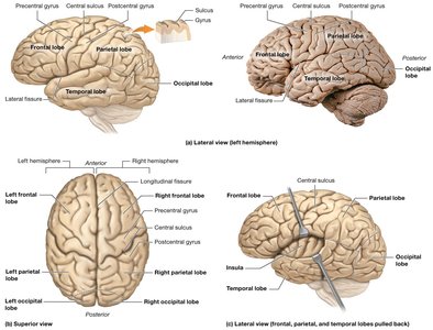
The cerebrum is divided into two hemispheres, each with four main lobes: frontal, parietal, temporal, and occipital. The insula is a fifth lobe located deep within the lateral fissure. The surface is marked by gyri (ridges), sulci (shallow grooves), and fissures (deep grooves).
Cerebral Cortex and Functional Areas
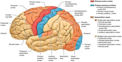
The cerebral cortex (neocortex) is the outer layer of gray matter, responsible for conscious processes. It contains primary motor and sensory cortices, as well as association areas for integration.
Primary Motor Cortex: Plans and executes movement.
Primary Sensory Cortices: Process sensory input (e.g., somatosensory, visual, auditory).
Association Areas: Integrate and interpret sensory and motor information.
Somatotopy and Homunculi
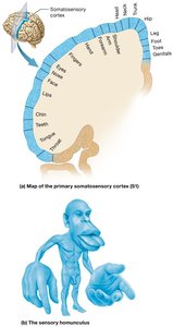
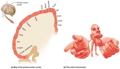
Specific regions of the cortex correspond to different body parts, depicted as sensory and motor homunculi. These maps illustrate the disproportionate representation of areas like the hands and face.
Basal Nuclei
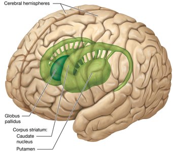
Basal nuclei are clusters of gray matter deep within the cerebral hemispheres. They regulate voluntary movement and inhibit involuntary movement. Major components include the caudate nucleus, putamen, and globus pallidus.
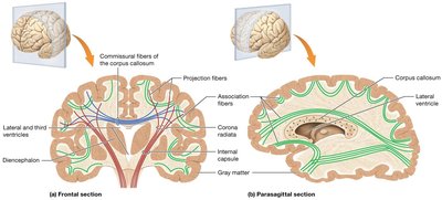
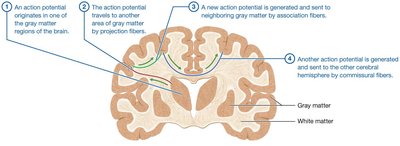
Cerebral White Matter
White matter tracts connect different regions of the brain:
Commissural Fibers: Connect right and left hemispheres (e.g., corpus callosum).
Projection Fibers: Connect cortex with lower brain regions and spinal cord.
Association Fibers: Connect regions within the same hemisphere.
Limbic System
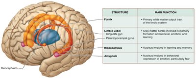
The limbic system is involved in emotion, memory, and behavior. Key structures include the limbic lobe, hippocampus (memory formation), amygdala (emotion, especially fear), and the fornix (major output tract).
The Diencephalon
Major Components
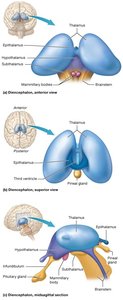
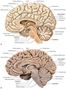
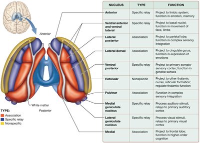
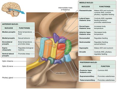
The diencephalon is located at the center of the brain and includes:
Thalamus: Relay station for sensory information.
Hypothalamus: Regulates homeostasis, endocrine functions, and the autonomic nervous system.
Epithalamus: Contains the pineal gland (secretes melatonin).
Subthalamus: Involved in movement control.
The Cerebellum
Structure and Function
The cerebellum is located posterior and inferior to the cerebrum. It coordinates voluntary movements, maintains posture and balance, and helps reduce motor error. It consists of two hemispheres connected by the vermis, with an outer cortex of gray matter and inner white matter (arbor vitae).
The Brainstem
Major Divisions
The brainstem connects the brain to the spinal cord and controls vital functions such as heart rate, breathing, and reflexes. It consists of the midbrain, pons, and medulla oblongata.
Summary Table: Major Brain Structures and Functions
Brain Division | Function |
|---|---|
Cerebrum | Performs higher mental functions, integrates sensory stimuli, plans and initiates movement |
Diencephalon | Processes, integrates, and relays information; maintains homeostasis; regulates biological rhythms |
Cerebellum | Monitors and coordinates movement |
Brainstem | Maintains homeostasis; controls certain reflexes; monitors movement; integrates and relays information |
Additional info:
The images included are directly relevant to the structure and function of the CNS as described in the text.
For more detailed study, refer to the specific sections on the spinal cord, protection of the CNS, and higher-order brain functions such as cognition, language, and memory.
