Back
BackThe Central Nervous System: Structure, Protection, and Function
Study Guide - Smart Notes
 Tailored notes based on your materials, expanded with key definitions, examples, and context.
The Central Nervous System (CNS)
Overview of CNS Functions
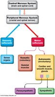
The central nervous system (CNS) is responsible for integrating sensory information, making decisions, and coordinating motor output. It consists of the brain and spinal cord, which together process and relay information throughout the body.
Sensory Functions: Detection of sensations both inside and outside the body.
Integrative Functions: Decision-making processes, including analysis and interpretation of sensory input.
Motor Functions: Stimulation of muscle contractions and glandular secretions.
Peripheral Nervous System (PNS): Performs sensory and motor functions, while integrative functions are exclusive to the CNS.
Basic Structure of the Brain
Major Regions of the Brain
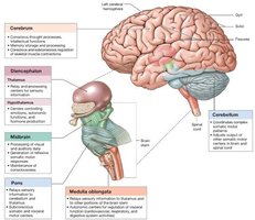
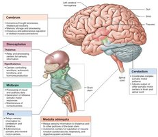
The adult human brain contains nearly 97% of the body's neural tissue and is divided into several major regions, each with specialized functions.
Cerebrum: Largest part; interprets sensory information and controls higher mental functions. Divided into right and left hemispheres by the longitudinal fissure. The surface is covered by the cerebral cortex (gray matter) with folds (gyri), shallow depressions (sulci), and deep grooves (fissures).
Cerebellum: Second largest; coordinates movement, evaluates sensory input, and is involved in timekeeping. Divided into hemispheres by the vermis and covered by the cerebellar cortex. The white matter forms the arbor vitae.
Brainstem: Relays information between the spinal cord and higher brain regions; controls basic life-sustaining functions. Includes the midbrain, pons, and medulla oblongata. The diencephalon (thalamus, hypothalamus, epithalamus) is functionally associated with the brainstem.
Gray Matter and White Matter
The brain consists of gray matter (neuron cell bodies, dendrites, and synapses) and white matter (myelinated axons). The cerebral cortex and basal nuclei are gray matter, while white matter lies deeper and connects different brain regions.
Protection and Support of the Brain
Physical Protection
Bones of the Cranium: Provide a rigid protective case.
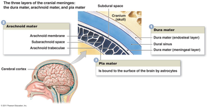
Cranial Meninges: Three connective tissue layers (dura mater, arachnoid mater, pia mater) that surround and protect the brain.
Cerebrospinal Fluid (CSF): Cushions the brain, provides buoyancy, and transports nutrients and waste.
Cranial Meninges
The meninges are three layers of connective tissue that protect the brain and spinal cord:
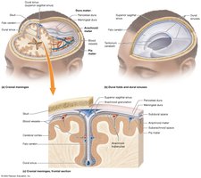
Dura Mater: Outermost, tough, and fibrous. Has periosteal (outer) and meningeal (inner) layers. Venous sinuses between layers drain blood from the brain.
Arachnoid Mater: Middle, smooth layer that does not dip into brain crevices. The subarachnoid space beneath contains CSF.
Pia Mater: Innermost, delicate layer that closely follows the brain's surface and enters sulci.
Dural Folds
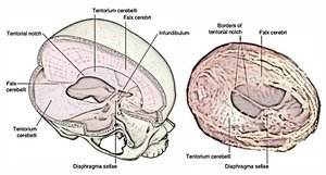
Folds of the dura mater extend into brain fissures to stabilize and support the brain:
Falx cerebri: Between cerebral hemispheres
Tentorium cerebelli: Separates cerebellum and cerebrum
Falx cerebelli: Divides cerebellar hemispheres
Cerebrospinal Fluid (CSF)
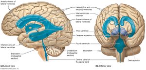
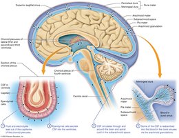
CSF is a clear fluid that circulates through the ventricles and around the CNS, providing cushioning, buoyancy, and nutrient transport.
Composition: Water, ions, glucose, some white blood cells, little protein, and wastes.
Functions: Cushions neural structures, provides buoyancy, transports nutrients and waste.
CSF Production and Flow
Produced by filtration of plasma through capillaries and ependymal cells in the choroid plexus.
Flows through the ventricles, exits via apertures, bathes the brain and spinal cord, and is reabsorbed into venous sinuses via arachnoid granulations.
About 500 mL produced daily; constantly drained into the bloodstream.
Blood Supply and Barriers
Blood Supply: Delivered by internal carotid and vertebral arteries; removed by internal jugular and vertebral veins. The brain consumes 20% of the body's oxygen and glucose.
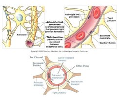
Blood-Brain Barrier (BBB): Formed by tight junctions between endothelial cells, restricting passage of substances. Lipid-soluble molecules diffuse; water and ions require channels; larger molecules use active transport. Astrocytes regulate permeability.
Blood-CSF Barrier: Specialized ependymal cells with tight junctions in the choroid plexus limit pathogen entry into CSF.
Breaks in BBB: Occur in the hypothalamus, pituitary gland, pineal gland, and choroid plexus for hormone secretion and CSF production.
Major Brain Regions and Their Functions
Cerebrum
The cerebrum is the largest brain region, responsible for conscious thought, intellectual functions, and voluntary motor activity. It is divided into lobes by sulci and fissures.
Lobes: Frontal, parietal, temporal, occipital, and insula.
Gray Matter: Outer cerebral cortex and basal nuclei.
White Matter: Association fibers (within hemisphere), commissural fibers (between hemispheres, e.g., corpus callosum), and projection fibers (to/from lower CNS).
Functional Areas of the Cerebral Cortex
Primary Motor Cortex: Precentral gyrus; initiates voluntary movement.
Premotor Cortex: Plans movements.
Primary Somatosensory Cortex: Postcentral gyrus; receives sensory input (touch, pressure, pain, temperature).
Association Areas: Interpret sensory input and plan responses.
Special Sensory Cortices: Visual (occipital), auditory (temporal), olfactory (medial temporal), gustatory (insula).
Prefrontal Cortex: Personality, decision-making, planning.
Wernicke’s Area: Language comprehension (usually left hemisphere).
Broca’s Area: Speech production (usually left hemisphere).
Limbic System
The limbic system is a functional grouping involved in emotion, motivation, and memory. It includes the limbic lobe, amygdala, hippocampus, thalamus, hypothalamus, and reticular formation.
Functions: Establishes emotional states, motivation, memory storage and retrieval.
Hippocampus: Organizes new memories and transfers them to the cerebral cortex for long-term storage.
Diencephalon
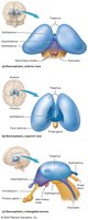
The diencephalon links the cerebrum with the brainstem and consists of the thalamus, hypothalamus, and epithalamus (including the pineal gland).
Thalamus: Relays and processes sensory information; part of the limbic system.
Hypothalamus: Major control center for the endocrine and autonomic nervous systems; regulates hormone production, thermoregulation, hunger, thirst, and emotions.
Pituitary Gland: Major endocrine gland controlled by the hypothalamus via the infundibulum.
Brainstem
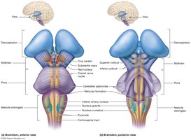
The brainstem connects the cerebrum and cerebellum to the spinal cord and is essential for basic life functions.
Midbrain (Mesencephalon): Visual and auditory reflexes, motor output, consciousness regulation.
Pons: Relays signals between cerebellum and brainstem; involved in sensory and motor functions, sleep, and respiration.
Medulla Oblongata: Regulates autonomic functions (heart rate, blood pressure, digestion); contains cardiac, vasomotor, and respiratory centers.
Cerebellum
The cerebellum coordinates voluntary movements, maintains posture and balance, and adjusts motor output based on sensory feedback.
Folia: Folds of the cerebellar cortex.
Arbor Vitae: Internal white matter.
Purkinje Cells: Large neurons in the cerebellar cortex.
Functions: Compares intended movement with actual performance and adjusts as needed (motor learning).
Spinal Cord Anatomy
External Structure
The spinal cord is located within the vertebral cavity, extending from the foramen magnum to the L1/L2 vertebrae. It serves as a major reflex center and pathway between the brain and peripheral nerves.
Conus Medullaris: Tapered end of the spinal cord.
Filum Terminale: Filamentous continuation anchoring the cord.
Cauda Equina: Bundle of lumbar, sacral, and coccygeal nerve roots.
Spinal Nerves and Meninges
31 pairs of spinal nerves: Formed by merging anterior (motor) and posterior (sensory) roots.
Meninges: Dura mater (outer), arachnoid mater (middle), pia mater (inner). The epidural space is present only in the spine.
Internal Structure
Gray Matter: Butterfly-shaped; contains anterior (motor), posterior (sensory), and lateral (autonomic) horns.
White Matter: Organized into ascending (sensory) and descending (motor) tracts.
Central Canal: Contains CSF.
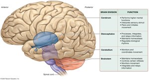
Summary Table: Major Brain Divisions and Functions
Brain Division | Function |
|---|---|
Cerebrum | Performs higher mental functions, interprets sensory stimuli, plans and initiates movement |
Diencephalon | Processes, integrates, and relays information; maintains homeostasis; regulates biological rhythms |
Cerebellum | Monitors and coordinates movement |
Brainstem | Maintains homeostasis, controls certain reflexes, monitors movement, integrates and relays information |