Back
BackThe Cranial Nerves: Structure, Function, and Classification
Study Guide - Smart Notes
 Tailored notes based on your materials, expanded with key definitions, examples, and context.
The Cranial Nerves
Overview of the Peripheral Nervous System: Cranial Nerves
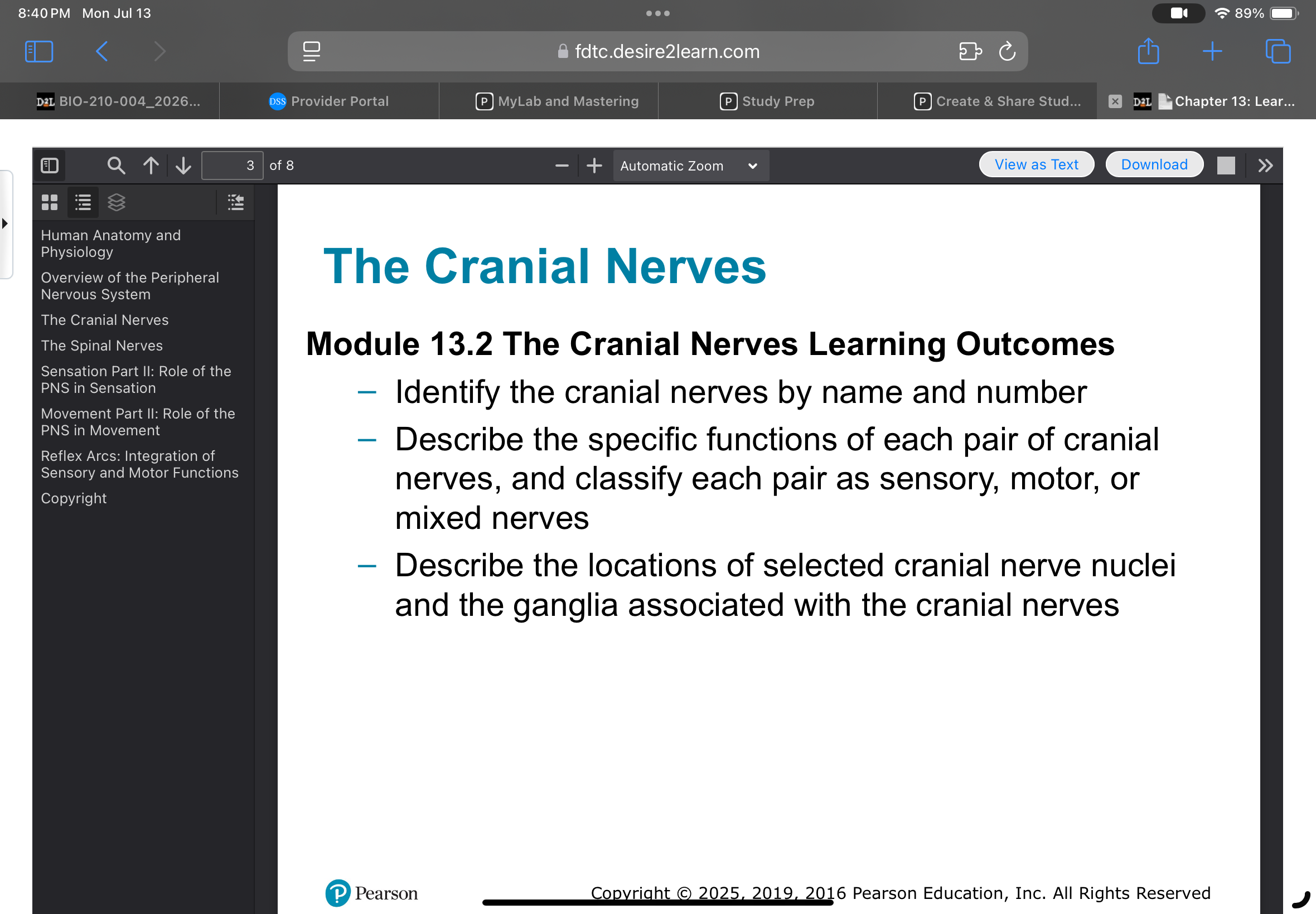
The cranial nerves are a set of twelve paired nerves that arise directly from the brain and brainstem, playing essential roles in sensory and motor functions of the head and neck. Understanding their names, numbers, functions, and anatomical locations is fundamental for students of human anatomy and physiology.
Identification of Cranial Nerves by Name and Number
Cranial nerves are traditionally numbered I through XII, based on their order from anterior to posterior.
Each nerve has a specific name reflecting its function or anatomical pathway.
Example: Olfactory nerve (I) is responsible for the sense of smell.
Classification and Functions of Cranial Nerves
Cranial nerves are classified as sensory, motor, or mixed, depending on the types of fibers they contain and their primary functions.
Sensory nerves transmit sensory information (e.g., smell, vision, hearing).
Motor nerves control muscle movements (e.g., eye movement, facial expression).
Mixed nerves contain both sensory and motor fibers (e.g., trigeminal nerve).
Each pair of cranial nerves has a unique set of functions, such as controlling eye movement, facial sensation, or swallowing.
Example: Facial nerve (VII) is a mixed nerve involved in facial expression and taste sensation.
Locations of Cranial Nerve Nuclei and Associated Ganglia
The nuclei of cranial nerves are located within specific regions of the brainstem, and some nerves are associated with ganglia that serve as relay points for sensory or motor signals.
Nuclei are clusters of neuron cell bodies in the central nervous system where cranial nerve fibers originate or terminate.
Ganglia are collections of neuron cell bodies in the peripheral nervous system, often associated with sensory cranial nerves.
Example: The trigeminal ganglion is associated with the trigeminal nerve (V), serving as a sensory relay.
Summary Table: Cranial Nerve Classification
Name | Number | Type | Main Function |
|---|---|---|---|
Olfactory | I | Sensory | Smell |
Optic | II | Sensory | Vision |
Oculomotor | III | Motor | Eye movement |
Trigeminal | V | Mixed | Facial sensation, chewing |
Facial | VII | Mixed | Facial expression, taste |
Vagus | X | Mixed | Autonomic control of heart, lungs, digestive tract |
Example: Clinical Relevance
Damage to the optic nerve (II) can result in vision loss.
Lesions affecting the facial nerve (VII) may cause facial paralysis (Bell's palsy).