 Back
BackThe Digestive System: Structure and Function
Study Guide - Smart Notes
 Tailored notes based on your materials, expanded with key definitions, examples, and context.
The Digestive System
Overview and Major Divisions
The digestive system is responsible for breaking down food, absorbing nutrients, and eliminating waste. It consists of the alimentary canal (gastrointestinal tract) and accessory digestive organs. The alimentary canal includes the mouth, pharynx, esophagus, stomach, small intestine, and large intestine. Accessory organs include the teeth, tongue, salivary glands, liver, gallbladder, and pancreas.
Alimentary canal: Continuous muscular tube from mouth to anus.
Accessory organs: Aid in digestion by producing secretions or mechanically processing food.
Major Processes of Digestion
Ingestion: Taking food into the mouth.
Propulsion: Moving food through the digestive tract (includes swallowing and peristalsis).
Mechanical digestion: Physical breakdown of food (chewing, churning in stomach, segmentation in intestines).
Chemical digestion: Enzymatic breakdown of food molecules into their building blocks.
Absorption: Transport of digested nutrients from the lumen of the GI tract into the blood or lymph.
Defecation: Elimination of indigestible substances as feces.
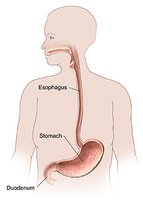
The Stomach
Gross Anatomy of the Stomach
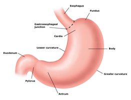
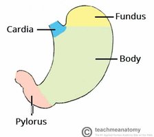
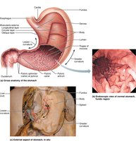
The stomach is a J-shaped organ located in the upper left quadrant of the abdomen. It acts as a temporary storage tank where food is churned and mixed with gastric juices to form chyme. The stomach initiates protein digestion and functions under highly acidic conditions.
Regions of the stomach: Cardia, fundus, body, pyloric part (pyloric antrum, pyloric canal, pyloric sphincter).
Curvatures: Lesser curvature (medial), greater curvature (lateral).
Rugae: Longitudinal folds in the mucosa that allow the stomach to expand as it fills.
Microscopic Anatomy of the Stomach
The stomach wall consists of four main layers: mucosa, submucosa, muscularis externa, and serosa. The mucosa is lined with simple columnar epithelium and contains gastric pits that open into gastric glands. The muscularis externa is unique in the stomach because it has three layers: circular, longitudinal, and oblique, which aid in churning food.
Mucosa: Secretes mucus to protect the stomach lining; contains gastric pits and glands.
Gastric glands: Contain several cell types:
Mucous neck cells: Secrete a special mucus.
Parietal cells: Secrete hydrochloric acid (HCl) and intrinsic factor.
Chief cells: Secrete pepsinogen (inactive enzyme, converted to pepsin by acid).
Enteroendocrine cells: Secrete hormones that regulate digestion.
Undifferentiated stem cells: Replace the epithelial lining every 3-7 days.
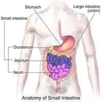
The Small Intestine
Gross Anatomy and Subdivisions
The small intestine is the longest part of the alimentary canal and is the primary site for enzymatic digestion and nutrient absorption. It is divided into three regions: duodenum, jejunum, and ileum.
Duodenum: Receives chyme from the stomach, digestive enzymes from the pancreas, and bile from the liver/gallbladder.
Jejunum: Middle section, major site of nutrient absorption.
Ileum: Final section, ends at the ileocecal valve (junction with the large intestine).
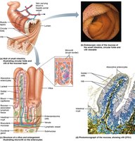
Microscopic Anatomy and Absorptive Adaptations
The small intestine is highly specialized for absorption, with three main structural modifications that increase surface area: circular folds, villi, and microvilli (the brush border).
Circular folds: Transverse ridges of mucosa and submucosa that slow the movement of chyme.
Villi: Fingerlike projections of the mucosa, each containing a capillary bed and a lymphatic vessel (lacteal) for nutrient absorption.
Microvilli: Tiny projections on the apical surface of absorptive cells, further increasing surface area.
Cell Types in the Small Intestine
Absorptive enterocytes: Uptake digested nutrients.
Goblet cells: Secrete mucus to lubricate chyme.
Enteroendocrine cells: Secrete hormones that regulate digestion.
The Large Intestine
Gross Anatomy and Subdivisions
The large intestine absorbs water and electrolytes from indigestible food residues and compacts them into feces. It is subdivided into the cecum, appendix, colon (ascending, transverse, descending, sigmoid), rectum, and anal canal.
Cecum: Initial pouch, receives material from the ileum.
Appendix: Contains lymphoid tissue, may serve as a reservoir for beneficial bacteria.
Colon: Four segments—ascending, transverse, descending, sigmoid.
Rectum and anal canal: Terminal portions; anal canal contains internal (involuntary) and external (voluntary) sphincters.

Special Features of the Large Intestine
Teniae coli: Three bands of longitudinal smooth muscle.
Haustra: Pouches formed by the contraction of the teniae coli.
Epiploic appendages: Fat-filled pouches of visceral peritoneum.
Microscopic Anatomy of the Large Intestine
Colonocytes: Absorptive cells for water and electrolytes.
Goblet cells: Secrete mucus to facilitate fecal movement.
Epithelium: Simple columnar, transitions to stratified squamous in the anal canal.
Villi: Absent in the large intestine.
Accessory Digestive Organs
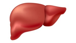
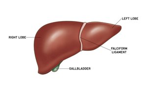
The Liver
The liver is the largest gland in the body and performs over 500 functions, including bile production for fat emulsification and numerous metabolic processes. It is divided into four lobes: right, left, caudate, and quadrate, separated by the falciform ligament.
Porta hepatis: Entry/exit point for vessels, nerves, and ducts.
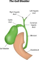
Hepatic ducts: Right and left hepatic ducts fuse to form the common hepatic duct.
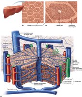
Microscopic Anatomy of the Liver
Liver lobules: Hexagonal units made of plates of hepatocytes radiating from a central vein.
Portal triad: Located at each lobule corner; consists of a bile duct, branch of the hepatic portal vein, and branch of the hepatic artery.
Bile canaliculi: Small ducts between hepatocytes that collect bile and drain into bile ducts.
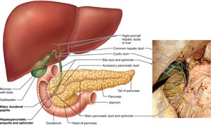
The Gallbladder
The gallbladder is a muscular sac that stores and concentrates bile produced by the liver. It releases bile into the duodenum via the cystic duct, which joins the common hepatic duct to form the bile duct. Bile is released in response to fatty food in the duodenum.
Mucosal folds: Allow expansion and contraction as bile is stored or released.
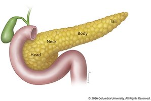
The Pancreas
The pancreas has both endocrine and exocrine functions. Its endocrine portion (islets of Langerhans) secretes insulin and glucagon to regulate blood sugar. The exocrine portion (acinar cells) produces digestive enzymes that are released into the duodenum via the main pancreatic duct.
Main pancreatic duct: Joins the bile duct to empty into the duodenum at the major duodenal papilla.
Acinar cells: Produce digestive enzymes (exocrine function).
Pancreatic islets: Secrete hormones (endocrine function).
Summary Table: Digestive Tract Regions and Functions
Region | Main Function(s) | Key Features |
|---|---|---|
Stomach | Churns food, begins protein digestion, forms chyme | Rugae, gastric glands, three muscle layers |
Small Intestine | Enzymatic digestion, nutrient absorption | Circular folds, villi, microvilli |
Large Intestine | Absorbs water/electrolytes, forms feces | Teniae coli, haustra, goblet cells |
Liver | Produces bile, metabolic functions | Lobules, portal triads, hepatocytes |
Gallbladder | Stores/concentrates bile | Mucosal folds, cystic duct |
Pancreas | Produces digestive enzymes, regulates blood sugar | Acinar cells, islets of Langerhans |
