 Back
BackThe Digestive System: Structure and Function
Study Guide - Smart Notes
 Tailored notes based on your materials, expanded with key definitions, examples, and context.
The Digestive System: Structure and Function
Overview of the Digestive System
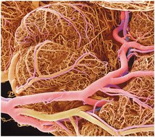
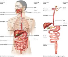
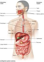
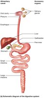
The digestive system is responsible for the breakdown of food, absorption of nutrients, and elimination of waste. It consists of the alimentary canal (gastrointestinal tract) and accessory organs that aid in digestion. The extensive blood supply to the digestive organs, especially the small intestine, facilitates rapid absorption of nutrients into the bloodstream.
Alimentary Canal: A continuous muscular tube that includes the mouth, pharynx, esophagus, stomach, small intestine, and large intestine.
Accessory Organs: Teeth, tongue, salivary glands, liver, gallbladder, and pancreas, which assist in the digestive process.
Main Functions: Ingestion, propulsion, mechanical digestion, chemical digestion, absorption, and defecation.
Blood Supply: The digestive organs are richly supplied with blood vessels to support nutrient absorption and transport.
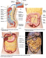
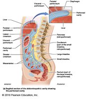
The Abdominopelvic Cavity and the Peritoneum
The digestive organs are located within the abdominopelvic cavity, which is lined by the peritoneum—the largest serous membrane in the body. The peritoneum supports the organs, provides a frictionless surface, and contains blood vessels, nerves, and lymphatics.
Parietal Peritoneum: Lines the internal surface of the abdominopelvic wall.
Visceral Peritoneum: Covers the external surfaces of most digestive organs.
Peritoneal Cavity: The space between the parietal and visceral layers, containing serous fluid.
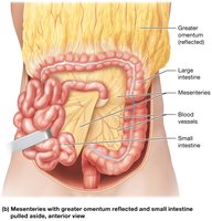
Mesenteries: Double layers of peritoneum that support and stabilize the intestines and contain blood vessels and nerves.
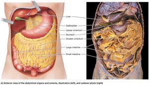
Omenta: Specialized folds of peritoneum (greater and lesser omentum) that protect and insulate abdominal organs.
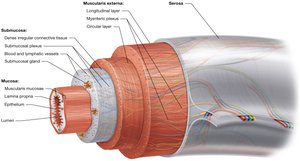
Histological Organization of the Alimentary Canal
The walls of the alimentary canal share a common structural organization, consisting of four main layers. These layers facilitate the movement, digestion, and absorption of food.
Mucosa: Innermost layer; consists of epithelium, lamina propria, and muscularis mucosae. Functions in secretion and absorption.
Submucosa: Dense connective tissue containing blood and lymphatic vessels, nerves, and glands.
Muscularis Externa: Two layers of smooth muscle (circular and longitudinal) responsible for peristalsis and segmentation.
Serosa (or Adventitia): Outermost layer; serosa is present in intraperitoneal organs, while adventitia is found in retroperitoneal organs.
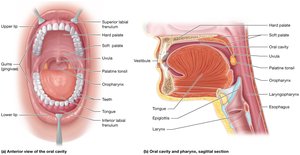
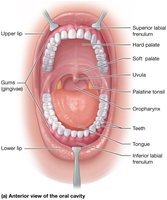
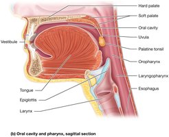
The Oral Cavity and Pharynx
The oral cavity (mouth) is the entry point for food and is involved in mechanical digestion (chewing) and chemical digestion (saliva). The pharynx serves as a passageway for food, fluids, and air.
Structures of the Oral Cavity: Lips, cheeks, palate (hard and soft), tongue, teeth, and salivary glands.
Pharynx: Divided into nasopharynx, oropharynx, and laryngopharynx; only the oropharynx and laryngopharynx are involved in digestion.
Function: Initiates swallowing (deglutition) and moves food into the esophagus.
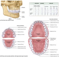
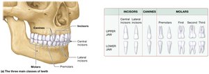
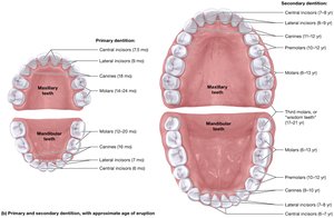
Teeth and Dentition
Teeth are essential for mechanical digestion, breaking food into smaller pieces to increase surface area for enzymes. Humans have two sets of teeth: primary (deciduous) and secondary (permanent) dentition.
Types of Teeth: Incisors (cutting), canines (tearing), premolars and molars (grinding).
Primary Dentition: 20 teeth, erupt between 6 months and 2 years of age.
Secondary Dentition: 32 teeth, replace primary teeth between ages 6 and 12 years.
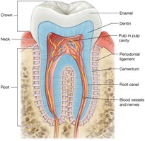
Structure of a Tooth
Each tooth consists of three main regions: crown, neck, and root. The internal structure supports its function in mastication (chewing).
Enamel: Hard, outermost layer; protects the tooth from abrasion and acid.
Dentin: Bone-like material beneath the enamel; forms the bulk of the tooth.
Pulp Cavity: Contains blood vessels and nerves; supplies nutrients and sensation.
Root: Anchors the tooth in the jawbone via the periodontal ligament.
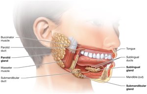
Salivary Glands
Salivary glands secrete saliva, which contains enzymes (such as amylase) that initiate the chemical digestion of carbohydrates and lubricate food for swallowing.
Major Salivary Glands: Parotid, submandibular, and sublingual glands.
Functions of Saliva: Moistens food, begins starch digestion, cleanses the mouth, and dissolves food chemicals for taste.
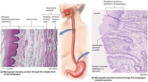
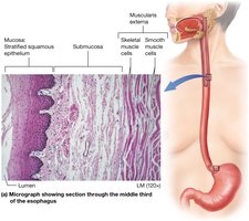
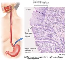
Histology of the Esophagus
The esophagus is a muscular tube that transports food from the pharynx to the stomach. Its wall structure is adapted for protection and propulsion of food.
Mucosa: Stratified squamous epithelium protects against abrasion.
Muscularis Externa: Upper third is skeletal muscle, middle third is mixed, and lower third is smooth muscle.
Adventitia: Outermost connective tissue layer anchors the esophagus to surrounding structures.
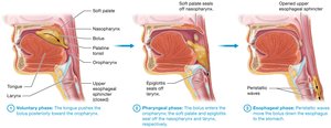
Swallowing (Deglutition)
Swallowing is a complex process involving voluntary and involuntary muscle contractions that move food from the mouth to the stomach.
Phases of Swallowing:
Voluntary Phase: Tongue pushes bolus toward the oropharynx.
Pharyngeal Phase: Involuntary; soft palate and epiglottis close off the nasopharynx and larynx, respectively.
Esophageal Phase: Peristaltic waves move the bolus down the esophagus.
 *Additional info: The notes above are expanded with academic context to ensure completeness and clarity for ANP college students studying the digestive system.*
