 Back
BackLecture 10.1: The Digestive System
Study Guide - Smart Notes
 Tailored notes based on your materials, expanded with key definitions, examples, and context.
The Digestive System
Overview and Major Functions
The digestive system is responsible for the breakdown of food, absorption of nutrients, and elimination of waste. It consists of the digestive tract (gastrointestinal tract) and accessory organs that aid in digestion through mechanical and chemical processes.
Ingestion: Intake of food through the oral cavity.
Mechanical digestion and propulsion: Physical breakdown and movement of food along the tract.
Chemical digestion: Enzymatic breakdown of food into absorbable molecules.
Secretion: Release of water, acids, enzymes, and buffers.
Absorption: Movement of nutrients, water, and electrolytes into the body.
Defecation: Elimination of indigestible substances as feces.
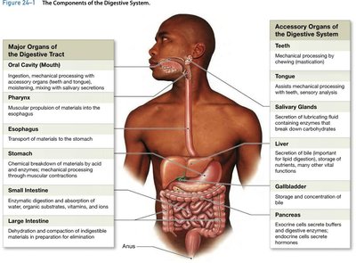
Organs of the Digestive System
Major Organs and Accessory Structures
The digestive tract is a continuous muscular tube extending from the oral cavity to the anus. Accessory organs include the teeth, tongue, salivary glands, liver, gallbladder, and pancreas, each contributing to the digestive process.
Oral Cavity (Mouth): Ingestion, mechanical processing, and mixing with salivary secretions.
Pharynx: Muscular propulsion of food into the esophagus.
Esophagus: Transport of materials to the stomach.
Stomach: Chemical breakdown and mechanical processing of food.
Small Intestine: Enzymatic digestion and absorption of nutrients.
Large Intestine: Dehydration, compaction, and elimination of indigestible materials.
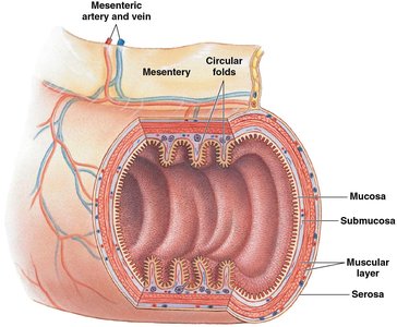
Histology of the Digestive Tract
Layers of the Digestive Tract
The wall of the digestive tract is composed of four major layers, each with specialized functions:
Mucosa: Innermost layer; contains epithelium, lamina propria, and muscularis mucosae.
Submucosa: Dense connective tissue with blood vessels, lymphatics, and nerves.
Muscular Layer: Smooth muscle responsible for peristalsis and segmentation.
Serosa: Outermost layer; a serous membrane covering most of the digestive tract.
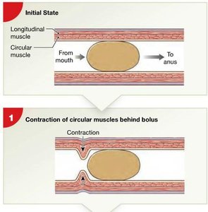
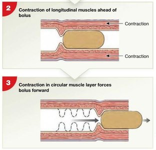
Motility: Peristalsis and Segmentation
Peristalsis
Peristalsis is a series of coordinated, wave-like muscle contractions that move food (bolus) through the digestive tract. It involves the contraction of circular and longitudinal muscles in a specific sequence:
Circular muscles behind the bolus contract, while those ahead relax.
Longitudinal muscles ahead of the bolus contract, shortening the tract segment.
A wave of contraction in the circular muscle layer pushes the bolus forward.
Deglutition (Swallowing)
Phases of Swallowing
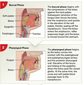
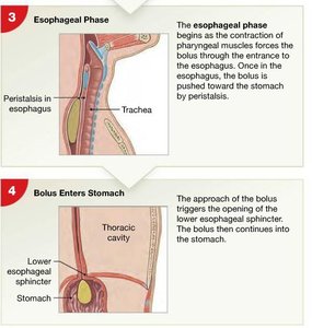
Swallowing is a complex process that moves food from the mouth to the stomach. It consists of three phases:
Buccal Phase: Voluntary; compression of the bolus against the hard palate and movement into the oropharynx.
Pharyngeal Phase: Involuntary; bolus contacts the pharyngeal wall, triggering reflexes that close the nasopharynx and larynx.
Esophageal Phase: Involuntary; peristalsis moves the bolus through the esophagus to the stomach.
The Stomach
Structure and Function
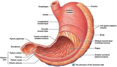
The stomach is a muscular, J-shaped organ that serves as a temporary storage site for food, initiates protein digestion, and mixes food with gastric secretions to form chyme.
Regions: Cardia, fundus, body, and pyloric part.
Curvatures: Lesser (medial) and greater (lateral).
Rugae: Folds in the mucosa that allow expansion.
Regulation of Gastric Activity
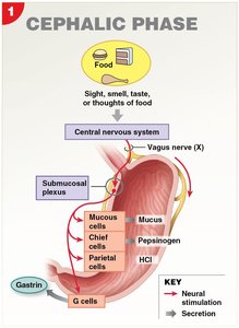
Gastric activity is regulated by neural, hormonal, and local mechanisms, occurring in three overlapping phases:
Cephalic Phase: Initiated by sight, smell, taste, or thought of food; stimulates gastric secretions via the vagus nerve.
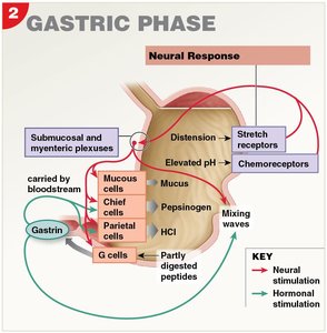
Gastric Phase: Begins with arrival of food in the stomach; stretch and chemoreceptors trigger secretions and mixing waves.
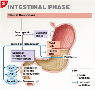
Intestinal Phase: Begins when chyme enters the duodenum; inhibits gastric activity and stimulates intestinal functions.
The Small Intestine
Structure and Segments
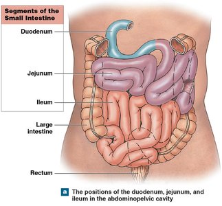
The small intestine is the primary site for chemical digestion and nutrient absorption. It is divided into three segments:
Duodenum: First 25 cm; receives chyme and digestive secretions from the pancreas and liver.
Jejunum: Middle 2.5 m; main site of digestion and absorption.
Ileum: Final 3.5 m; ends at the ileocecal valve, controlling entry into the large intestine.
Accessory Digestive Organs
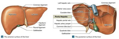
Pancreas
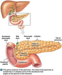
The pancreas is both an endocrine and exocrine organ. It lies posterior to the stomach and is divided into head, body, and tail regions. The pancreatic duct delivers digestive enzymes and bicarbonate to the duodenum.
Endocrine function: Secretion of insulin and glucagon to regulate blood glucose.
Exocrine function: Secretion of pancreatic juice containing digestive enzymes and bicarbonate.
Liver
The liver is the largest visceral organ and performs metabolic, hematological, and synthetic functions. It is divided into lobes and lobules, which are the functional units.
Metabolic regulation: Carbohydrate, lipid, and amino acid metabolism; detoxification; vitamin and mineral storage.
Hematological regulation: Removal of old red blood cells, synthesis of plasma proteins, and detoxification of blood.
Bile production: Bile emulsifies fats, aiding in their digestion and absorption.
The Large Intestine
Structure and Function
The large intestine absorbs water and electrolytes, compacts indigestible materials, and stores feces prior to defecation. It consists of the cecum, colon, and rectum.
Cecum: Pouch-like first portion.
Colon: Largest portion, subdivided into ascending, transverse, descending, and sigmoid regions.
Rectum: Final 15 cm; stores feces until elimination.
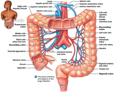
Functions of the Large Intestine
Absorption of water, bile salts, vitamins, and some nutrients.
Compaction of intestinal contents into feces.
Storage of fecal material prior to defecation.
Host to a diverse microbiome that synthesizes vitamins (e.g., vitamin K, biotin, and vitamin B5).
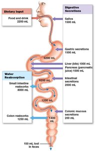
Water Movement in the Digestive System
Absorption and Secretion
Water movement in the digestive tract is passive and follows osmotic gradients. Most water absorption occurs in the small intestine, with additional reabsorption in the colon.
Integration with Other Body Systems
Systemic Interactions
The digestive system interacts with multiple organ systems to maintain homeostasis. For example, it provides nutrients to all body systems, while the cardiovascular system transports absorbed nutrients, and the nervous and endocrine systems regulate digestive activity.
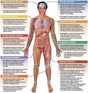
Additional info: The digestive system's efficiency and health are critical for overall metabolism, immunity, and energy balance. Disorders of the digestive tract can impact nutrient status and systemic health.
