 Back
The Digestive System: Structure, Function, and Clinical Correlations
Study Guide - Smart Notes
 Tailored notes based on your materials, expanded with key definitions, examples, and context.
The Digestive System
Overview of the Digestive System
The digestive system is responsible for the breakdown of food, absorption of nutrients, and elimination of waste. It consists of the alimentary canal and accessory digestive organs. The alimentary canal includes the mouth, pharynx, esophagus, stomach, small intestine, and large intestine. Accessory organs such as the teeth, tongue, salivary glands, liver, gallbladder, and pancreas assist in digestion by producing secretions that facilitate the breakdown of food.
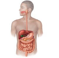
Alimentary canal: Continuous muscular tube from mouth to anus.
Accessory organs: Connected by ducts; their secretions aid digestion.
Digestive Processes
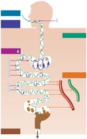
Digestion involves several coordinated processes that move and chemically break down food, absorb nutrients, and eliminate waste.
Ingestion: Taking food into the mouth.
Propulsion: Movement of food through the alimentary canal, primarily by peristalsis.
Mechanical breakdown: Physical preparation of food for chemical digestion (chewing, churning, segmentation).
Digestion: Enzymatic breakdown of complex molecules into absorbable units.
Absorption: Transport of digested nutrients into blood or lymph.
Defecation: Elimination of indigestible substances as feces.
Motility: Peristalsis and Segmentation
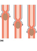
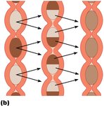
Motility in the digestive tract is essential for moving and mixing food. Two primary types of movements are peristalsis and segmentation.
Peristalsis: Involuntary, wave-like contractions of smooth muscle that propel food forward through the digestive tract.
Segmentation: Rhythmic contractions that mix food with digestive juices and increase contact with the mucosa for absorption.
Histology of the Alimentary Canal
General Structure
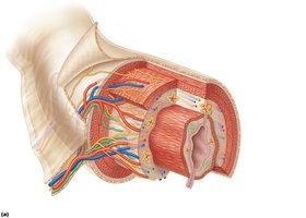
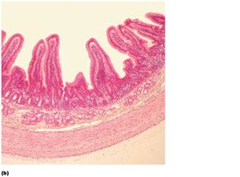
The wall of the alimentary canal from the esophagus to the anus is composed of four main layers:
Mucosa: Innermost layer; consists of epithelium, lamina propria, and muscularis mucosae.
Submucosa: External to mucosa; contains blood and lymphatic vessels, nerves, and glands.
Muscularis externa: Responsible for peristalsis and segmentation; usually has an inner circular and outer longitudinal layer.
Serosa: Outermost layer; the visceral peritoneum.
Smooth Muscle in the Digestive Tract
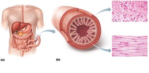
Smooth muscle is the primary muscle type in the walls of the digestive organs, allowing for involuntary movements such as peristalsis and segmentation.
Structure: Elongated fibers with a single central nucleus, organized into sheets.
Layers: Longitudinal (outer) and circular (inner) layers.
The Mouth and Associated Organs
Anatomy of the Mouth
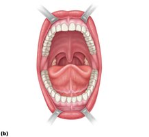
The mouth, or oral cavity, is the entry point for food and is lined by a mucosal layer of stratified squamous epithelium. Its boundaries include the lips, cheeks, palate, and tongue.
Lips and cheeks: Formed by the orbicularis oris and buccinator muscles.
Palate: Forms the roof of the mouth; separates oral and nasal cavities.
Fauces: Opening to the oropharynx, bounded by palatoglossal and palatopharyngeal arches.
The Tongue
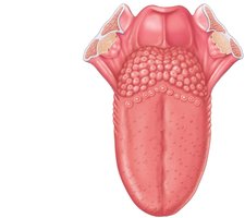
The tongue is a muscular organ that manipulates food, assists in swallowing, and is involved in speech. It contains both intrinsic and extrinsic muscles and is anchored by the lingual frenulum.
Papillae: Projections on the superior surface; filiform (no taste buds), fungiform, and vallate (contain taste buds).
Sulcus terminalis: Divides the anterior two-thirds from the posterior one-third of the tongue.
The Teeth
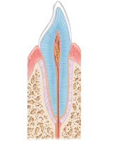
Teeth are essential for mechanical digestion. Humans have two sets: deciduous (milk) teeth and permanent teeth. The dental formula for adults is 2I, 1C, 2P, 3M (incisors, canines, premolars, molars).
Structure: Crown (exposed), neck, and root (in socket).
Enamel: Hard outer layer; dentin lies beneath enamel; pulp cavity contains nerves and blood vessels.
The Salivary Glands
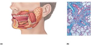
Salivary glands produce saliva, which contains enzymes that initiate carbohydrate digestion and mucus for lubrication. Major glands include the parotid, submandibular, and sublingual glands.
Parotid gland: Located near the ear; secretes via the parotid duct.
Submandibular gland: Medial to the mandible.
Sublingual gland: Floor of the mouth; mainly mucous cells.
The Pharynx and Esophagus
The Pharynx
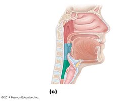
The pharynx serves as a passageway for both air and food. The oropharynx and laryngopharynx are lined with stratified squamous epithelium and have an external muscle layer composed of pharyngeal constrictors.
The Esophagus
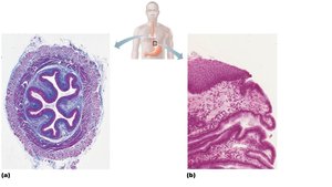
The esophagus is a muscular tube that transports food from the pharynx to the stomach. It begins at the continuation of the pharynx and ends at the stomach, passing through the diaphragm. The cardiac sphincter prevents reflux of stomach acid.
Microscopic anatomy: Lined with nonkeratinized stratified squamous epithelium; mucosa and submucosa form longitudinal folds when empty.
Muscularis externa: Upper third is skeletal muscle, middle third is mixed, lower third is smooth muscle.
Adventitia: Outermost connective tissue layer.
The Stomach
Gross Anatomy
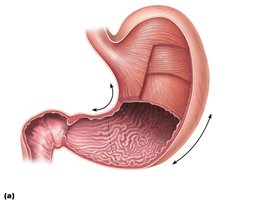
The stomach is a muscular organ where food is churned into chyme and protein digestion begins. It functions under acidic conditions and typically retains food for about four hours. Major regions include the cardia, fundus, body, and pyloric part.
Microscopic Anatomy
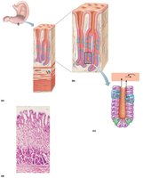
The stomach wall has three layers of muscularis externa (longitudinal, circular, and oblique). The mucosa is lined with simple columnar epithelium and contains gastric pits leading to gastric glands.
Mucous neck cells: Secrete mucus.
Parietal cells: Secrete hydrochloric acid and intrinsic factor.
Chief cells: Secrete pepsinogen, which is activated to pepsin in acidic conditions.
Enteroendocrine cells: Secrete hormones.
The Small Intestine
Gross Anatomy
The small intestine is the longest part of the alimentary canal and is the primary site for enzymatic digestion and nutrient absorption. It is divided into three regions: duodenum, jejunum, and ileum.
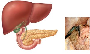
Duodenum: Receives bile and pancreatic enzymes via ducts.
Jejunum and ileum: Main sites for absorption.
Microscopic Anatomy
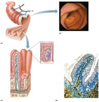
The small intestine is highly specialized for absorption, with several modifications to increase surface area:
Circular folds (plicae circulares): Transverse ridges of mucosa and submucosa.
Villi: Fingerlike projections of the mucosa covered with simple columnar epithelium.
Microvilli: Extensions of the plasma membrane of absorptive cells, forming the brush border.
Cell types: Absorptive enterocytes (nutrient uptake), goblet cells (mucus secretion), enteroendocrine cells (hormone secretion), and cells in intestinal crypts (intestinal juice secretion).
The Large Intestine
Gross Anatomy
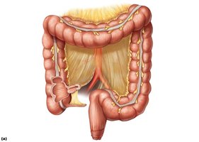
The large intestine absorbs water and electrolytes from indigestible food residues and compacts them into feces. It consists of the cecum, appendix, colon (ascending, transverse, descending, sigmoid), rectum, and anal canal.
Special features: Teniae coli (longitudinal muscle bands), haustra (pouches), and epiploic appendages (fat-filled pouches).
Microscopic Anatomy
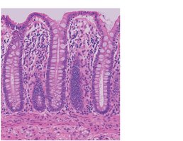
The large intestine lacks villi but contains numerous goblet cells and simple tubular glands called intestinal crypts. The epithelium transitions to stratified squamous in the anal canal.
Accessory Digestive Organs
The Liver
The liver is the largest gland in the body and performs over 500 functions, including bile production for fat emulsification and various metabolic processes.
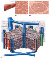
Microscopic structure: Organized into lobules with hepatocytes, portal triads (bile duct, portal vein, hepatic artery), and sinusoids containing stellate macrophages.
Functions of hepatocytes: Protein synthesis, bile production, detoxification, storage of glycogen, and regeneration.
The Gallbladder
The gallbladder stores and concentrates bile produced by the liver and releases it into the duodenum in response to cholecystokinin, which is secreted when fatty chyme enters the small intestine. Bile aids in the emulsification of fats.
The Pancreas
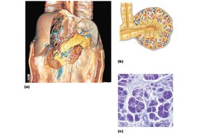
The pancreas has both endocrine and exocrine functions. Endocrine cells produce insulin and glucagon to regulate blood sugar, while exocrine acinar cells produce digestive enzymes that are released into the small intestine.
Main pancreatic duct: Joins the bile duct to form the hepatopancreatic ampulla, which empties into the duodenum.
Blood supply: Hepatic, splenic, and superior mesenteric arteries.
Clinical Correlations and Disorders
Peptic Ulcers
Peptic ulcers are erosions of the mucosa in the stomach (gastric ulcers) or duodenum (duodenal ulcers). They are primarily caused by infection with Helicobacter pylori, which induces acid overproduction and inflammation.
Symptoms: Abdominal pain, nausea, and possible bleeding.
Treatment: Antibiotics, acid-suppressing medications.
Other Digestive Disorders
Intestinal obstruction: Can be mechanical (adhesions, tumors) or nonmechanical (paralysis of peristalsis).
Inflammatory bowel disease: Includes Crohn’s disease and ulcerative colitis, characterized by chronic inflammation of the intestinal wall.
Viral hepatitis: Inflammation of the liver, often presenting with jaundice and flu-like symptoms; types include A, B, C, and G.
Cystic fibrosis: Thick mucus blocks pancreatic ducts, leading to malabsorption of nutrients.
