 Back
BackThe Digestive System: Structure, Function, and Processes
Study Guide - Smart Notes
 Tailored notes based on your materials, expanded with key definitions, examples, and context.
The Digestive System
Function of the Digestive System
The digestive system is responsible for the intake, breakdown, absorption, and elimination of food. Its main functions include:
Ingestion: Taking in food through the mouth.
Digestion: Breaking down food into nutrient molecules by mechanical and chemical means.
Absorption: Moving nutrient molecules into the bloodstream for distribution throughout the body.
Elimination: Ridding the body of indigestible remains as feces.
Overview of Digestive System Anatomy
The organs of the digestive system are divided into two groups: the alimentary canal and accessory digestive organs.
Alimentary canal (GI tract): A continuous muscular tube running from the mouth to the anus, including the mouth, pharynx, esophagus, stomach, small intestine, large intestine, and anus. It digests and absorbs food.
Accessory digestive organs: Teeth, tongue, gallbladder, and digestive glands (salivary glands, liver, pancreas) that assist in the breakdown of food.
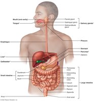
Digestive Processes
Food is processed through several coordinated steps:
Ingestion: Eating food.
Propulsion: Moving food through the alimentary canal, including swallowing and peristalsis (waves of muscle contraction and relaxation).
Mechanical breakdown: Chewing, mixing with saliva, churning in the stomach, and segmentation in the intestines.
Digestion: Enzymatic breakdown of complex molecules into simpler building blocks.
Absorption: Passage of digested fragments into blood or lymph.
Defecation: Elimination of indigestible substances as feces.
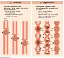
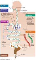
Organization and Histology of the Digestive System
Relationship of Digestive Organs to the Peritoneum
The peritoneum is a serous membrane lining the abdominal cavity and covering abdominal organs. It consists of:
Visceral peritoneum: Covers external surfaces of most digestive organs.
Parietal peritoneum: Lines the body wall.
Peritoneal cavity: Fluid-filled space between the two peritoneums, allowing organs to move smoothly.
Mesentery: Double layer of peritoneum that supports organs, provides routes for blood vessels, lymphatics, and nerves, and stores fat.
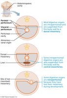
Histology of the Alimentary Canal
All digestive organs share four basic layers (tunics):
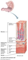
Mucosa: Innermost layer; secretes mucus, digestive enzymes, and hormones; absorbs end products; protects against infection.
Submucosa: Areolar connective tissue with blood and lymphatic vessels, lymphoid follicles, and nerves.
Muscularis externa: Responsible for segmentation and peristalsis; has inner circular and outer longitudinal muscle layers.
Serosa: Outermost layer; visceral peritoneum.
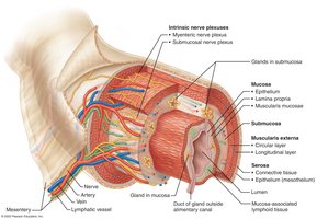
Control of Digestive System
Enteric Nervous System
The GI tract has its own nervous system, the enteric nervous system (ENS), also called the "gut brain." It contains more neurons than the spinal cord and regulates digestive tract motility and secretion.
Submucosal nerve plexus: Regulates glands and smooth muscle in the mucosa.
Myenteric nerve plexus: Controls GI tract motility.
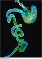
Regulation of Digestive Activity
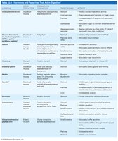
Digestive activity is regulated by mechanical and chemical stimuli, neural (intrinsic and extrinsic) controls, and hormones. Short reflexes are mediated by the ENS, while long reflexes involve the central nervous system. Parasympathetic stimulation enhances digestion, while sympathetic stimulation inhibits it.
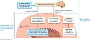
Functional Anatomy of the Digestive System
Mouth and Associated Organs
The mouth (oral cavity) is where food is chewed and mixed with saliva, beginning the process of digestion. Associated organs include the tongue, salivary glands, and teeth.
Lips and cheeks: Form the boundaries of the mouth; help keep food between teeth during chewing.
Palate: Forms the roof of the mouth; hard palate is bony, soft palate is muscular and closes off the nasopharynx during swallowing.
Uvula: Prevents food from entering the nasal cavity.
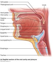
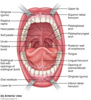
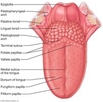
Tongue
The tongue grips, repositions, and mixes food during chewing, forms the bolus, and initiates swallowing, speech, and taste. It contains various types of papillae, some of which house taste buds.
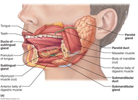
Salivary Glands
Salivary glands produce saliva, which cleanses the mouth, dissolves food chemicals, moistens food, and begins the breakdown of starch with amylase. Major glands include the parotid, submandibular, and sublingual glands.
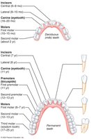
The Teeth
Teeth are essential for mastication (chewing), which mechanically breaks food into smaller fragments. Humans have two sets of teeth: primary (deciduous) and permanent. Teeth are classified as incisors, canines, premolars, and molars based on their shape and function.
Pharynx and Esophagus
The Pharynx
The pharynx allows passage of food, fluids, and air. It is lined with stratified squamous epithelium and has skeletal muscle layers for swallowing.
The Esophagus
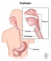
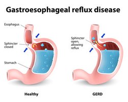
The esophagus is a muscular tube that transports food from the pharynx to the stomach. The gastroesophageal (cardiac) sphincter prevents acid reflux.
Clinical: Heartburn and GERD
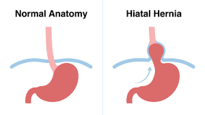
Heartburn is caused by stomach acid regurgitating into the esophagus, often due to a weak sphincter or hiatal hernia. Chronic reflux can lead to gastroesophageal reflux disease (GERD), esophagitis, ulcers, or cancer.
Deglutition (Swallowing)
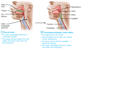
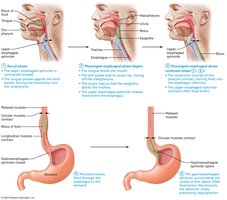
Swallowing involves a voluntary buccal phase and an involuntary pharyngeal-esophageal phase, coordinated by the swallowing center in the brainstem.
The Stomach
Gross Anatomy of the Stomach
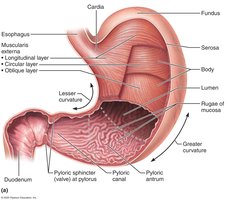
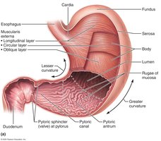
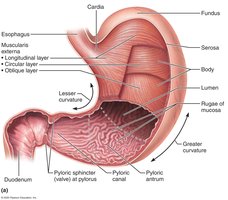
The stomach is a temporary storage tank that initiates protein digestion and converts food into chyme. It has four main regions: cardia, fundus, body, and pylorus. The stomach wall contains rugae (folds) when empty.
Microscopic Anatomy of the Stomach
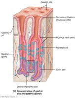
The stomach wall has three layers of smooth muscle (including an oblique layer) for churning food. The mucosa is lined with simple columnar epithelium and contains gastric pits leading to gastric glands, which produce gastric juice.
Types of Gland Cells
Mucous neck cells: Secrete thin, acidic mucus.
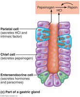
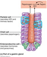
Parietal cells: Secrete hydrochloric acid (HCl) and intrinsic factor (for vitamin B12 absorption).
Chief cells: Secrete pepsinogen (activated to pepsin) and gastric lipase.
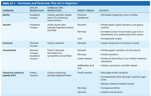
Enteroendocrine cells: Secrete hormones and paracrines (e.g., gastrin, histamine, somatostatin).
Mucosal Barrier
The stomach is protected from its acidic environment by a mucosal barrier consisting of a thick layer of bicarbonate-rich mucus, tight junctions between epithelial cells, and rapid cell turnover.
Clinical: Gastritis and Ulcers
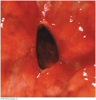
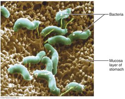
Gastritis is inflammation of the stomach mucosa, often caused by Helicobacter pylori infection or NSAIDs. Peptic ulcers are erosions of the stomach wall that can lead to serious complications.
Digestive Processes in the Stomach
Breakdown of food and mixing with gastric juice to form chyme.
Protein digestion by pepsin.
Absorption of some substances (e.g., alcohol, aspirin).
Secretion of intrinsic factor (essential for vitamin B12 absorption).
Regulation of Gastric Secretion
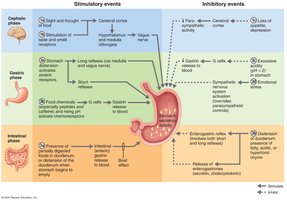
Gastric secretion is regulated by neural (vagus nerve, sympathetic inhibition) and hormonal (gastrin) mechanisms. There are three phases:
Cephalic phase: Triggered by sight, smell, taste, or thought of food.
Gastric phase: Triggered by food in the stomach; involves stretch and chemical receptors.
Intestinal phase: Brief stimulation as chyme enters the duodenum, followed by inhibition.
Liver, Gallbladder, and Pancreas
The Liver
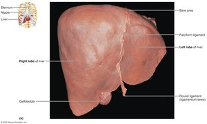
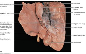
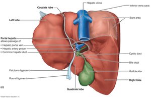
The liver is the largest gland in the body and produces bile, which emulsifies fats. It has four lobes and is supported by the falciform and round ligaments. The porta hepatis is the entry/exit point for blood vessels and bile ducts.
Liver Microscopic Anatomy
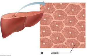
Liver lobules are hexagonal units made of hepatocytes. Each lobule has a central vein and portal triads (branch of hepatic artery, portal vein, and bile duct). Blood flows through sinusoids, where nutrients are processed and toxins removed.
The Gallbladder
The gallbladder stores and concentrates bile. Gallstones can form if there is too much cholesterol or too few bile salts, leading to pain and possible jaundice.
The Pancreas
The pancreas produces digestive enzymes (pancreatic juice) and bicarbonate to neutralize stomach acid. It also has endocrine functions (insulin and glucagon secretion).
The Small Intestine
Gross Anatomy
The small intestine is the major organ of digestion and absorption, divided into the duodenum, jejunum, and ileum. It receives blood from the superior mesenteric artery and is innervated by both parasympathetic and sympathetic nerves.
Microscopic Anatomy
The small intestine has structural modifications to increase surface area for absorption:
Circular folds: Permanent folds that force chyme to spiral through the lumen.
Villi: Fingerlike projections containing capillaries and lacteals for nutrient absorption.
Microvilli: Cytoplasmic extensions forming the brush border, which contains digestive enzymes.
The Large Intestine
Gross Anatomy
The large intestine absorbs water and electrolytes, forms feces, and eliminates waste. It includes the cecum, appendix, colon (ascending, transverse, descending, sigmoid), rectum, and anal canal. Unique features include teniae coli, haustra, and epiploic appendages.
Bacterial Flora
The large intestine houses a diverse bacterial community that ferments indigestible carbohydrates, synthesizes vitamins, and helps keep pathogenic bacteria in check.
Digestive Processes in the Large Intestine
Residue remains in the large intestine for 12–24 hours. Major functions are propulsion of feces and defecation. No significant food breakdown occurs except by bacteria.
Physiology of Digestion and Absorption
Mechanism of Digestion: Enzymatic Hydrolysis
Digestion is a catabolic process that breaks macromolecules into monomers small enough for absorption, primarily through enzymatic hydrolysis.
Mechanisms of Absorption
Absorption involves moving substances from the gut lumen into the body, primarily through epithelial cells by active or passive transport. Most nutrients are absorbed before chyme reaches the ileum.
Digestion and Absorption of Nutrients
Carbohydrates: Broken down to monosaccharides (glucose, fructose, galactose) by amylases and brush border enzymes.
Proteins: Digested to amino acids by pepsin (stomach) and pancreatic/brush border proteases.
Lipids: Emulsified by bile salts, digested by pancreatic lipases, absorbed as fatty acids and monoglycerides, reassembled into triglycerides, and transported as chylomicrons.
Nucleic acids: Broken down by pancreatic nucleases and brush border enzymes into nitrogenous bases, pentose sugars, and phosphate ions.
Absorption of Vitamins, Electrolytes, and Water
Fat-soluble vitamins (A, D, E, K): Absorbed with micelles.
Water-soluble vitamins (C, B): Absorbed by diffusion or active transport.
Electrolytes: Actively absorbed along the small intestine; iron and calcium absorption is regulated by need.
Water: Absorbed by osmosis, mainly in the small intestine.
Clinical Correlations
Lactose intolerance: Deficiency of lactase leads to inability to digest lactose, causing diarrhea and gas.
Malabsorption diseases: Can result from interference with bile or pancreatic juice delivery, or damage to intestinal mucosa (e.g., celiac disease).
