 Back
BackThe Digestive System: Structure, Function, and Physiology
Study Guide - Smart Notes
 Tailored notes based on your materials, expanded with key definitions, examples, and context.
The Digestive System
Overview and Main Functions
The digestive system is responsible for the breakdown of food, absorption of nutrients, and elimination of indigestible substances. It consists of the alimentary canal and accessory digestive organs, each playing a specific role in the digestive process.
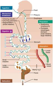
Ingestion: Taking in food through the mouth.
Mechanical Breakdown: Chewing, mixing, and churning food to increase surface area for enzymes.
Propulsion: Movement of food through the alimentary canal via swallowing and peristalsis.
Chemical Digestion: Enzymatic breakdown of complex food molecules into their chemical building blocks.
Absorption: Passage of digested nutrients from the GI tract into the blood or lymph.
Defecation: Elimination of indigestible substances as feces.
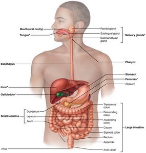
Organs of the Digestive System
Alimentary Canal (GI Tract): A continuous muscular tube from mouth to anus, including the mouth, pharynx, esophagus, stomach, small intestine, large intestine, and anus. It digests and absorbs food.
Accessory Digestive Organs: Teeth, tongue, gallbladder, salivary glands, liver, and pancreas. These organs produce secretions that aid in digestion.
Digestive Processes
Six Essential Activities
Ingestion
Propulsion (swallowing and peristalsis)
Mechanical Breakdown (chewing, churning, segmentation)
Chemical Digestion
Absorption
Defecation
Peristalsis is the major means of propulsion, involving waves of contraction and relaxation. Segmentation is a local constriction that mixes food with digestive juices.

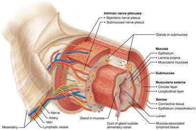
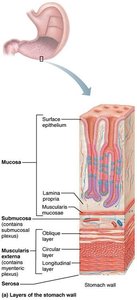
Histology and Structure of the Alimentary Canal
Basic Layers
The alimentary canal wall consists of four basic layers:
Mucosa: Innermost layer; secretes mucus, digestive enzymes, and hormones; absorbs end products; protects against disease.
Submucosa: Contains blood vessels, lymphatics, and nerves.
Muscularis Externa: Responsible for segmentation and peristalsis; contains inner circular and outer longitudinal muscle layers.
Serosa: Outermost layer; protective connective tissue.
Control of Digestive System
Enteric Nervous System
The GI tract has its own nervous system, the enteric nervous system (ENS), also known as the "gut brain." It contains more neurons than the spinal cord and regulates GI tract motility and secretion.
Short Reflexes: Mediated by the ENS; respond to local stimuli such as stretch, pH, and chemical composition.
Long Reflexes: Involve the CNS and autonomic nervous system; respond to stimuli inside or outside the gut.
Parasympathetic Input: Stimulates digestive activity.
Sympathetic Input: Inhibits digestive activity.
Digestive activity is also regulated by hormones released from cells in the stomach and small intestine.
Functional Anatomy of the Digestive System
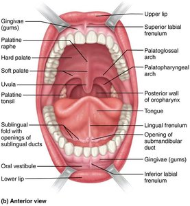
Mouth and Associated Organs
The mouth is the entry point for food, where it is chewed and mixed with saliva to begin digestion. Associated organs include the tongue, salivary glands, and teeth.
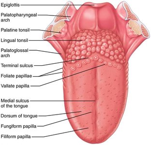
Tongue: Grips, repositions, and mixes food; forms the bolus; initiates swallowing, speech, and taste.
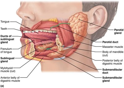
Salivary Glands: Produce saliva, which cleanses the mouth, dissolves food chemicals, moistens food, and begins starch digestion with amylase.
Teeth: Involved in mastication (chewing), breaking food into smaller fragments.
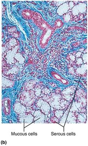
Salivary Glands
Serous Cells: Produce watery secretions with enzymes and ions.
Mucous Cells: Produce mucus.
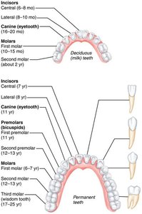
Teeth and Dentition
Primary Dentition: 20 deciduous (baby) teeth erupt between 6–24 months.
Permanent Dentition: 32 teeth, including incisors (cutting), canines (tearing), premolars and molars (grinding).
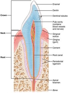
Tooth Structure
Crown: Exposed part above the gum, covered by enamel (hardest substance in the body).
Root: Embedded in the jawbone, covered by cement, attached by periodontal ligament.
Dentin: Bonelike material under enamel, maintained by odontoblasts.
Pulp Cavity: Contains connective tissue, blood vessels, and nerves.
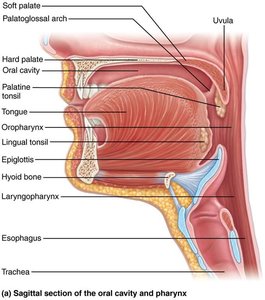
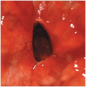
Pharynx and Esophagus
Pharynx
The pharynx allows passage of food, fluids, and air. It is lined with stratified squamous epithelium and contains skeletal muscle layers for swallowing.
Esophagus
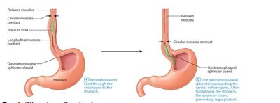
The esophagus is a muscular tube that transports food from the pharynx to the stomach. The gastroesophageal sphincter prevents acid reflux.
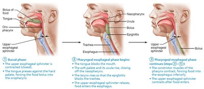
Deglutition (Swallowing)
Swallowing involves coordinated activity of over 22 muscle groups and includes the buccal phase (voluntary) and pharyngeal-esophageal phase (involuntary).
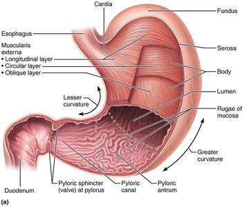
The Stomach
Gross Anatomy
The stomach is a temporary storage tank that initiates protein digestion and converts food into chyme. It can expand significantly due to rugae (folds in the mucosa).
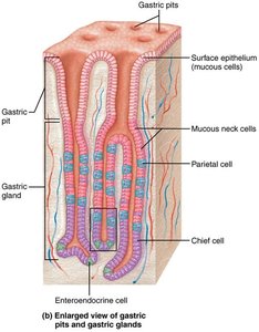
Microscopic Anatomy
Mucosa: Simple columnar epithelium with mucous cells; contains gastric pits leading to gastric glands.
Gastric Glands: Contain mucous neck cells, parietal cells (secrete HCl and intrinsic factor), chief cells (secrete pepsinogen), and enteroendocrine cells (secrete hormones).

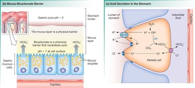
Mucosal Barrier
The stomach is protected from harsh acidic conditions by a mucosal barrier consisting of a thick layer of bicarbonate-rich mucus, tight junctions between epithelial cells, and rapid cell turnover.
Clinical Correlates
Gastritis: Inflammation due to breach of the mucosal barrier.
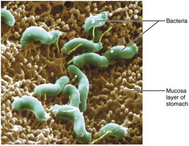
Peptic Ulcers: Erosions in the stomach wall, often caused by Helicobacter pylori infection or NSAIDs.
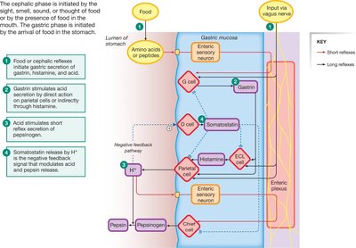
Regulation of Gastric Secretion
Cephalic Phase: Triggered by sight, smell, taste, or thought of food.
Gastric Phase: Initiated by food in the stomach; involves stretch and chemical stimuli.
Intestinal Phase: Begins with chyme entering the small intestine; initially stimulates, then inhibits gastric secretion.
Summary Table: Functions of Gastrointestinal Organs
Organ | Main Function(s) |
|---|---|
Mouth | Ingestion, mechanical breakdown, chemical digestion (carbohydrates) |
Stomach | Mechanical breakdown, chemical digestion (proteins), food storage, production of intrinsic factor |
Small Intestine | Major site of digestion and absorption |
Large Intestine | Absorption of water and electrolytes, formation and elimination of feces |
Liver | Production of bile, processing of nutrients |
Pancreas | Secretion of digestive enzymes and bicarbonate |
Gallbladder | Storage and concentration of bile |
Additional info: Table content inferred and summarized from the overview of gastrointestinal organ functions.
