 Back
BackThe Ear: Structure, Function, and Hearing Process
Study Guide - Smart Notes
 Tailored notes based on your materials, expanded with key definitions, examples, and context.
The Ear: Structure and Function
Overview of the Ear
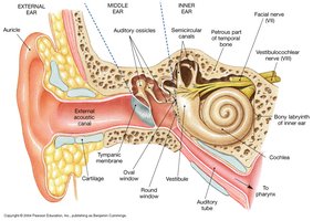
The ear is a complex organ responsible for hearing and balance. It is divided into three main regions: the external ear, middle ear, and inner ear. Each region has specialized structures that contribute to the processes of sound detection and equilibrium.
External Ear
Anatomy and Functions
Pinna (Auricle): The visible, fleshy, cartilaginous part that gathers sound waves and directs them into the ear canal.
External Acoustic Meatus (Canal): A passageway that channels sound waves toward the tympanic membrane.
Tympanic Membrane (Eardrum): A thin, semitransparent sheet that vibrates in response to sound waves, separating the external ear from the middle ear.
Ceruminous Glands: Located along the external acoustic meatus, these glands secrete cerumen (ear wax) to protect the ear by trapping debris and slowing microorganism growth.
Middle Ear
Structure and Function
Tympanic Cavity: An air-filled chamber within the petrous portion of the temporal bone.
Pharyngotympanic Tube (Eustachian Tube): Connects the middle ear to the pharynx, equalizing air pressure on both sides of the tympanic membrane.
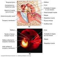
Auditory Ossicles: Three small bones that transmit and amplify vibrations from the tympanic membrane to the inner ear:
Malleus (Hammer): Attaches to the tympanic membrane.
Incus (Anvil): Connects the malleus to the stapes.
Stapes (Stirrup): Base attaches to the oval window of the inner ear.
Tensor Tympani Muscle: Inserts on the malleus to reduce movement under loud conditions; innervated by the trigeminal nerve (V).
Stapedius Muscle: Inserts on the stapes to reduce movement under loud conditions; innervated by the facial nerve (VII).
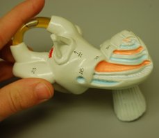
Inner Ear
Major Components and Functions
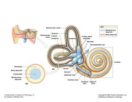
Semicircular Canals: Detect rotational movements of the head and contribute to equilibrium.
Vestibule: Senses gravitational pull and linear acceleration; contains the utricle and saccule.
Cochlea: Spiral-shaped chamber responsible for hearing.
Vestibulocochlear Nerve (VIII): Transmits sensory information from the inner ear to the brain.
Inner Ear Organization
Bony Labyrinth: Rigid, bony outer wall that protects the membranous labyrinth.
Membranous Labyrinth: Network of fluid-filled tubes within the bony labyrinth.
Perilymph: Fluid between the bony and membranous labyrinths.
Endolymph: Fluid within the membranous labyrinth.
Windows of the Inner Ear
Oval Window: Receives vibrations from the stapes, creating pressure waves in the vestibular duct.
Round Window: Allows dissipation of pressure waves from the tympanic duct, preventing echo within the cochlea.
Receptor Function in the Inner Ear
Hair Cells and Supporting Structures
Hair Cells: Mechanoreceptors sensitive to movement, responsible for detecting sound and head position.
Stereocilia: Long microvilli on the surface of hair cells that respond to mechanical stimuli.
Kinocilium: A single, large cilium that helps detect direction of movement.
Supporting Cells: Orient hair cells vertically and provide structural support.
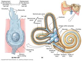
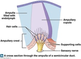
Semicircular Ducts and Ampullae
Ampulla: Expanded region at the base of each semicircular duct containing the ampullary crest (crista) with hair cells.
Cupula: Gelatinous structure bound to the crista, containing hair cells and extending across the ampulla.
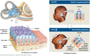
Vestibule: Maculae and Otolithic Membrane
Maculae: Oval structures in the saccule and utricle containing hair cells that detect linear acceleration and gravity.
Otolithic Membrane: Gelatinous layer covering hair cells, topped with otoliths (calcium carbonate granules) that enhance sensitivity to movement.
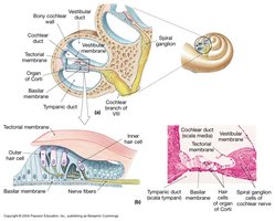
Cochlea and the Organ of Corti
Spiral Organ (Organ of Corti)
The Organ of Corti is the primary sensory structure for hearing, located within the cochlear duct between the vestibular and tympanic ducts. It contains hair cells that transduce mechanical vibrations into nerve impulses.
Vestibular Membrane: Separates cochlear duct from vestibular duct.
Basilar Membrane: Separates cochlear duct from tympanic duct and supports the Organ of Corti.
Tectorial Membrane: Overlies the hair cells' stereocilia in the Organ of Corti.
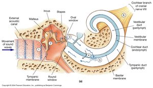
The Hearing Process
Steps in Hearing
Sound waves arrive at the tympanic membrane, causing it to vibrate.
Vibrations are transmitted through the auditory ossicles (malleus, incus, stapes).
Movement of the stapes at the oval window creates pressure waves in the perilymph of the vestibular duct.
Pressure waves distort the basilar membrane as they travel toward the round window of the tympanic duct.
Vibration of the basilar membrane causes hair cells to move against the tectorial membrane, generating nerve impulses.
Information about the region and intensity of stimulation is relayed to the central nervous system via the cochlear branch of the vestibulocochlear nerve (VIII).
Summary Table: Major Structures of the Ear
Region | Main Structures | Function |
|---|---|---|
External Ear | Pinna, External Acoustic Meatus, Tympanic Membrane, Ceruminous Glands | Collects sound, directs waves, protects ear |
Middle Ear | Tympanic Cavity, Ossicles (Malleus, Incus, Stapes), Tensor Tympani, Stapedius, Pharyngotympanic Tube | Amplifies and transmits sound, equalizes pressure |
Inner Ear | Semicircular Canals, Vestibule, Cochlea, Vestibulocochlear Nerve | Balance, equilibrium, hearing |
Additional info: The notes above expand on the provided lecture content with academic context, definitions, and logical organization for comprehensive study. Images were included only when directly relevant to the adjacent explanation.
