 Back
BackThe Ear: Structure, Function, and Mechanisms of Hearing and Equilibrium
Study Guide - Smart Notes
 Tailored notes based on your materials, expanded with key definitions, examples, and context.
The Ear: Structure and Function
Overview of the Ear
The ear is a complex organ responsible for detecting sound and maintaining balance. It is divided into three main regions: the outer ear, middle ear, and inner ear. Each region plays a specific role in the processes of hearing and equilibrium.
Sound: Physical Properties
Nature of Sound Waves
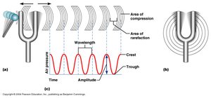
Sound is a pressure disturbance produced by a vibrating object, which creates a sound wave that travels through a medium such as air. The properties of sound waves determine how we perceive pitch and loudness.
Wavelength: The distance between two consecutive wave crests. Shorter wavelengths correspond to higher frequencies.
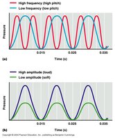
Frequency: The number of waves passing a point per second, measured in hertz (Hz). Higher frequency means higher pitch. Human hearing ranges from 20 to 20,000 Hz.
Amplitude: The height of the wave, which determines the loudness of the sound. Loudness is measured in decibels (dB).
Examples of Sound Levels
15 dB: Rustling leaves
45 dB: Normal conversation
60 dB: Crowd noise
75 dB: Vacuum cleaner
90 dB: Drill (frequent exposure can cause hearing loss)
115 dB: Pain threshold
120 dB: Loud rock music
Anatomy of the Ear
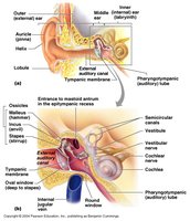
Outer Ear
The outer ear collects and channels sound waves toward the middle ear.
Pinna (Auricle): Elastic cartilage structure that catches sound waves.
Auditory Canal: Passageway that carries sound to the middle ear. Contains ceruminous glands that produce wax to trap bacteria and particles, providing a waterproof coating.
Middle Ear
The middle ear amplifies and transmits sound vibrations from the outer ear to the inner ear.
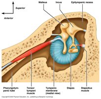
Tympanic Membrane (Eardrum): Thin, fibrous connective tissue that vibrates in response to sound waves.
Ossicles: Three small bones (malleus, incus, stapes) that transmit and amplify vibrations. The malleus is attached to the tympanic membrane, the incus connects the malleus to the stapes, and the stapes presses on the oval window of the inner ear.
Eustachian Tube: Equalizes air pressure on both sides of the tympanic membrane and drains the middle ear.
Inner Ear
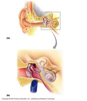
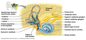
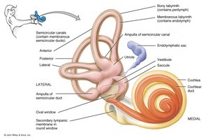
The inner ear contains structures for both hearing and balance, housed within the bony labyrinth.
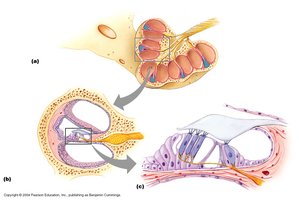
Cochlea: Spiral-shaped organ responsible for hearing.
Semicircular Canals: Three canals oriented in different planes, responsible for detecting head motion (dynamic equilibrium).
Saccule and Utricle: Detect the position of the head in space (static equilibrium).
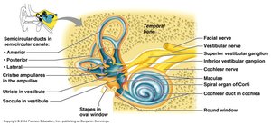
Cochlea: Structure and Function
The cochlea is divided into three ducts:
Vestibular Duct (Scala Vestibuli): Upper duct filled with perilymph.
Tympanic Duct (Scala Tympani): Lower duct filled with perilymph.
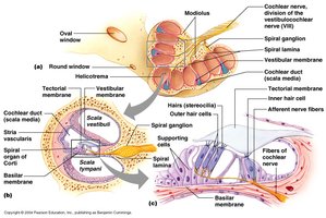
Cochlear Duct (Scala Media): Middle duct filled with endolymph, containing the Organ of Corti.
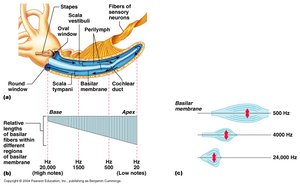
Organ of Corti
The Organ of Corti is the sensory organ of hearing, located within the cochlear duct.
Sensory Hair Cells: Rest on the basilar membrane and have stereocilia that contact the tectorial membrane.
Basilar Membrane: Vibrates in response to sound, causing hair cells to bend.
Tectorial Membrane: Stiff membrane that interacts with hair cell stereocilia.
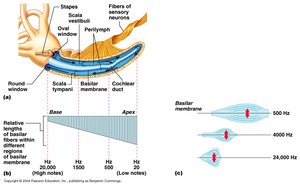
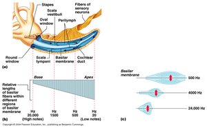
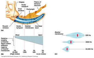
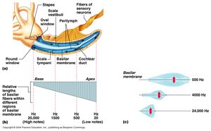
Frequency Mapping in the Cochlea
Different regions of the basilar membrane respond to different frequencies:
High Frequency: Detected at the base (near the stapes).
Low Frequency: Detected at the apex (helicotrema).
Mechanisms of Hearing
Pathway of Sound
Hearing involves the transmission of sound waves through the ear structures, resulting in nerve impulses sent to the brain.
Sound waves enter the auditory canal and strike the tympanic membrane, causing it to vibrate.
Vibrations are transmitted and amplified by the ossicles (malleus, incus, stapes).
The stapes moves the oval window, creating pressure waves in the perilymph of the vestibular duct.
Pressure waves travel through the cochlea, causing the basilar membrane to vibrate at specific locations depending on frequency.
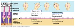
Vibration of the basilar membrane bends the hair cells against the tectorial membrane, generating nerve impulses in the vestibulocochlear nerve.
Equilibrium: Balance and Orientation
Semicircular Canals: Dynamic Equilibrium
The semicircular canals detect rotational movements of the head. Each canal is oriented in a different plane and contains fluid and sensory hair cells that respond to acceleration, deceleration, and rotation.
Saccule and Utricle: Static Equilibrium
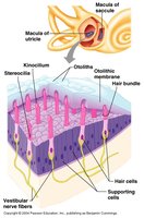
The saccule and utricle detect the position of the head relative to gravity. Hair cells are covered by a gelatinous layer with otoliths (calcium carbonate crystals). When the head tilts, otoliths shift, pulling on the hair cells and signaling changes in head position.
Summary Table: Key Structures and Functions of the Ear
Region | Main Structures | Function |
|---|---|---|
Outer Ear | Pinna, Auditory Canal | Collects and channels sound waves |
Middle Ear | Tympanic Membrane, Ossicles, Eustachian Tube | Amplifies and transmits sound; equalizes pressure |
Inner Ear | Cochlea, Semicircular Canals, Saccule, Utricle | Hearing (cochlea); balance and equilibrium (semicircular canals, saccule, utricle) |
Key Equations
Frequency (f): where is the period (time for one cycle).
Speed of Sound (v): where is the wavelength.
Decibel Level (dB): where is the intensity of the sound and is the reference intensity.
