 Back
BackThe Endocrine System and The Cardiovascular System: Heart Anatomy
Study Guide - Smart Notes
 Tailored notes based on your materials, expanded with key definitions, examples, and context.
The Endocrine System
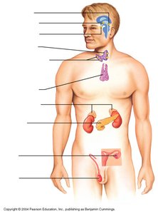
Overview of Major Endocrine Organs
The endocrine system is a collection of glands that produce hormones regulating metabolism, growth, development, tissue function, sexual function, reproduction, sleep, and mood. The organs are distributed throughout the body and release hormones directly into the bloodstream.
Pituitary Gland: Often called the 'master gland,' it regulates other endocrine glands and produces hormones such as growth hormone and ACTH.
Thyroid Gland: Located in the neck, it produces hormones that regulate metabolism, such as thyroxine (T4) and triiodothyronine (T3).
Parathyroid Glands: Small glands behind the thyroid, they regulate calcium levels in the blood.
Adrenal Glands: Located above the kidneys, they produce hormones like cortisol and adrenaline, which help the body respond to stress.
Pancreas: Produces insulin and glucagon, which regulate blood glucose levels.
Testes: Produce testosterone, which regulates male reproductive development and function.
Example: The adrenal glands release adrenaline during stressful situations, preparing the body for 'fight or flight.'
The Cardiovascular System: The Heart
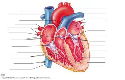
Internal Anatomy of the Heart
The heart is a muscular organ responsible for pumping blood throughout the body. It consists of four chambers: two atria and two ventricles. The internal anatomy includes valves, septa, and major vessels that ensure unidirectional blood flow.
Atria: The upper chambers that receive blood from the body (right atrium) and lungs (left atrium).
Ventricles: The lower chambers that pump blood to the lungs (right ventricle) and the rest of the body (left ventricle).
Valves: Prevent backflow of blood. Includes the tricuspid, bicuspid (mitral), pulmonary, and aortic valves.
Septum: The wall dividing the right and left sides of the heart.
Major Vessels: Superior and inferior vena cava, pulmonary arteries and veins, and the aorta.
Example: The left ventricle pumps oxygenated blood into the aorta for systemic circulation.
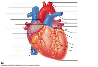
External Anatomy of the Heart
The external anatomy of the heart includes the coronary arteries and veins, which supply blood to the heart muscle itself. The heart is encased in the pericardium, a protective sac.
Coronary Arteries: Supply oxygen-rich blood to the heart muscle.
Coronary Veins: Remove deoxygenated blood from the heart muscle.
Pericardium: The double-walled sac that surrounds and protects the heart.
Example: Blockage of a coronary artery can lead to a myocardial infarction (heart attack).
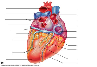
Major Vessels and Surface Features of the Heart
The heart's surface features include the major vessels entering and leaving the heart, as well as grooves and sulci that mark the boundaries between chambers.
Aorta: The largest artery, carries blood from the left ventricle to the body.
Pulmonary Arteries: Carry deoxygenated blood from the right ventricle to the lungs.
Pulmonary Veins: Return oxygenated blood from the lungs to the left atrium.
Vena Cavae: Superior and inferior, return deoxygenated blood from the body to the right atrium.
Grooves/Sulci: Mark the boundaries between the atria and ventricles and contain coronary vessels.
Example: The coronary sulcus separates the atria from the ventricles and houses the coronary arteries.
Summary Table: Heart Chambers and Valves
The following table summarizes the main chambers and valves of the heart:
Chamber | Function | Associated Valve |
|---|---|---|
Right Atrium | Receives deoxygenated blood from body | Tricuspid Valve |
Right Ventricle | Pumps blood to lungs | Pulmonary Valve |
Left Atrium | Receives oxygenated blood from lungs | Bicuspid (Mitral) Valve |
Left Ventricle | Pumps blood to body | Aortic Valve |
Key Equations
Cardiac output is a fundamental concept in cardiovascular physiology:
Cardiac Output (CO): The volume of blood pumped by the heart per minute.
Equation:
Where:
Stroke Volume (SV): The amount of blood pumped by the left ventricle in one contraction.
Heart Rate (HR): The number of heartbeats per minute.
Example: If stroke volume is 70 mL and heart rate is 75 beats/min, cardiac output is mL/min.
