 Back
BackThe Endocrine System: Hypothalamus and Pituitary Gland
Study Guide - Smart Notes
 Tailored notes based on your materials, expanded with key definitions, examples, and context.
The Endocrine System: Hypothalamus and Pituitary Gland
Overview of the Hypothalamus and Pituitary Gland
The hypothalamus and pituitary gland are central components of the endocrine system, responsible for regulating numerous physiological processes through hormone secretion. The hypothalamus connects to the pituitary gland via the infundibulum, a stalk-like structure. The pituitary gland consists of two major lobes: the posterior pituitary (neurohypophysis) and the anterior pituitary (adenohypophysis).
Hypothalamus: Located below the thalamus, it controls the pituitary gland.
Pituitary Gland: Secretes at least eight major hormones.
Infundibulum: The stalk connecting hypothalamus and pituitary.
Posterior Pituitary: Neural tissue, secretes neurohormones.
Anterior Pituitary: Glandular tissue, secretes peptide hormones.
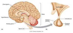
Anatomical Relationships and Imaging
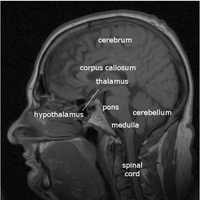
The pituitary gland is situated at the base of the brain, beneath the hypothalamus. MRI imaging is used to visualize the anatomical relationships between the hypothalamus, pituitary gland, and other brain structures.
Posterior lobe: Derived from neural tissue, maintains neural connection to hypothalamus.
Anterior lobe: Derived from oral mucosa, connected via blood vessels.
Posterior Pituitary and Hypothalamic Hormones
Neural Connections and Hormone Release
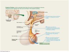
The posterior pituitary is composed of axon terminals from hypothalamic neurons. These neurons manufacture two key neurohormones: oxytocin and antidiuretic hormone (ADH). Hormones are stored in axon terminals and released into the bloodstream upon neuronal activation.
Paraventricular neurons: Produce oxytocin.
Supraoptic neurons: Produce ADH.
Both hormones are peptides, each composed of nine amino acids, differing by two amino acids.
Oxytocin
Oxytocin is a strong stimulant of uterine contractions during childbirth and acts as a hormonal trigger for milk ejection. It is released in response to stretching of the cervix and infant suckling, both positive feedback mechanisms. Oxytocin also acts as a neurotransmitter in the brain, promoting bonding and trust.
Uterine contractions: Facilitates labor.
Milk ejection: Targets myoepithelial cells for milk release.
Behavioral effects: Promotes nurturing and couple bonding.
Clinical use: Synthetic oxytocin is used to induce or hasten labor.
Antidiuretic Hormone (ADH)
ADH inhibits urine production by promoting water reabsorption in kidney tubules. Its release is triggered by high solute concentration, pain, low blood pressure, and certain drugs. Alcohol inhibits ADH, leading to increased urine output and dehydration. Severe blood loss stimulates ADH release, causing vasoconstriction and increased blood pressure (hence, ADH is also called vasopressin).
Osmoreceptors: Monitor solute concentration in hypothalamus.
Kidney tubules: Targeted for water reabsorption.
Vasoconstriction: ADH increases blood pressure during blood loss.
Inhibition: Alcohol and excess water intake inhibit ADH.
Anterior Pituitary and Hormone Regulation
Vascular Connections and Hormone Regulation
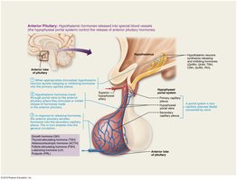
The anterior pituitary is glandular tissue connected to the hypothalamus via the hypophyseal portal system, which consists of two capillary plexuses and portal veins. The hypothalamus secretes releasing and inhibiting hormones to regulate anterior pituitary hormone secretion.
Primary capillary plexus
Hypophyseal portal veins
Secondary capillary plexus
Releasing/inhibiting hormones: Control hormone secretion.
Anterior Pituitary Hormones
The anterior pituitary secretes six peptide hormones, four of which are tropic hormones that regulate other endocrine glands. These hormones include:
Growth hormone (GH): Somatotropin, promotes growth and metabolism.
Thyroid-stimulating hormone (TSH): Tropic, stimulates thyroid gland.
Adrenocorticotropic hormone (ACTH): Tropic, stimulates adrenal cortex.
Follicle-stimulating hormone (FSH): Tropic, stimulates gamete production.
Luteinizing hormone (LH): Tropic, stimulates gonadal hormone production.
Prolactin (PRL): Stimulates milk production.
Growth Hormone (GH)
GH is an anabolic hormone that promotes tissue building and growth. It has glucose-sparing actions, stimulates glycogenolysis, increases fatty acid levels, and encourages protein synthesis. GH triggers the production of insulin-like growth factors (IGFs) in the liver, skeletal muscle, and bone, which stimulate cell division and bone matrix formation.
Metabolic effects: Decreases glucose uptake, increases fatty acid use.
Growth effects: Stimulates cell enlargement and division, especially in bone and muscle.
Regulation: Controlled by GHRH (stimulates) and GHIH (inhibits) from hypothalamus.
Equation: GH regulation can be summarized as:
Thyroid-Stimulating Hormone (TSH)
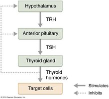
TSH is a tropic hormone produced by thyrotropic cells, stimulating the thyroid gland's development and activity. Its release is triggered by thyrotropin-releasing hormone (TRH) from the hypothalamus and inhibited by rising thyroid hormone levels and GHIH.
Stimulates: Thyroid gland to produce thyroid hormones.
Regulation: Negative feedback from thyroid hormones.
Adrenocorticotropic Hormone (ACTH)
ACTH stimulates the adrenal cortex to release corticosteroids. Its release is triggered by corticotropin-releasing hormone (CRH) from the hypothalamus, with highest levels in the morning. ACTH helps resist stressors, and its release is influenced by fever, hypoglycemia, and stress.
Stimulates: Adrenal cortex for corticosteroid production.
Regulation: Triggered by CRH, affected by stress and metabolic factors.
Gonadotropins (FSH and LH)
FSH and LH are secreted by gonadotropic cells and regulate reproductive functions. FSH stimulates gamete production, while LH promotes gonadal hormone production. In females, LH triggers ovulation and estrogen/progesterone release; in males, LH stimulates testosterone production.
Absent in prepubertal children.
Regulation: Triggered by GnRH during and after puberty; suppressed by gonadal hormones (negative feedback).
Prolactin (PRL)
Prolactin stimulates milk production in females and is regulated primarily by prolactin-inhibiting hormone (PIH), which is dopamine. PRL levels rise with estrogen and during pregnancy, and suckling stimulates further PRL release for continued milk production.
Milk production: Stimulated by PRL.
Regulation: Controlled by PIH (dopamine); decreased PIH leads to lactation.
Unique regulation: No negative feedback by PRL.
Summary Table: Anterior Pituitary Hormones
Hormone | Main Function | Regulation |
|---|---|---|
GH | Growth, metabolism | GHRH/GHIH from hypothalamus |
TSH | Stimulates thyroid | TRH from hypothalamus, negative feedback by thyroid hormones |
ACTH | Stimulates adrenal cortex | CRH from hypothalamus, stress factors |
FSH | Gamete production | GnRH from hypothalamus, negative feedback by gonadal hormones |
LH | Gonadal hormone production | GnRH from hypothalamus, negative feedback by gonadal hormones |
PRL | Milk production | PIH (dopamine) from hypothalamus |
Additional info: The hypothalamic-pituitary axis is a central regulatory system for endocrine function, integrating neural and hormonal signals to maintain homeostasis.
