 Back
BackThe Endocrine System: Mechanisms, Glands, and Hormonal Regulation
Study Guide - Smart Notes
 Tailored notes based on your materials, expanded with key definitions, examples, and context.
The Endocrine System and Its Comparison to the Nervous System
Modes of Communication
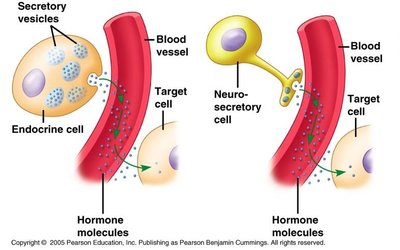
The endocrine system and nervous system are the two primary regulatory systems in the human body. They differ in their methods of communication, speed, and duration of effects.
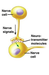
Nervous System: Utilizes electrical impulses and neurotransmitters for rapid, short-lived responses. Information is transmitted directly between nerve cells.
Endocrine System: Uses hormones released into the bloodstream, resulting in slower but longer-lasting effects.
Hormone Concentrations and Tissue Response
Hormone Half-life and Concentration
Hormones have a characteristic half-life, which is the time required for their plasma concentration to decrease by half. This can range from minutes to days. Most hormones are metabolized by the liver and excreted as less active products.
Physiological Concentrations: Tissues respond to hormones only within a normal range. Excessive (pharmacological) concentrations can cause abnormal effects, including cross-reactivity with other hormone receptors and side effects.
Priming and Desensitization
Priming (Up-regulation): Target cells may increase their receptor numbers in response to a hormone, making them more sensitive to future stimulation.
Desensitization (Down-regulation): Prolonged exposure to high hormone levels can decrease receptor numbers, reducing sensitivity. Many hormones are released in pulses to prevent this.
Mechanisms of Hormone Action
Hormone-Receptor Interactions
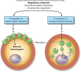
Hormones exert their effects by binding to specific receptors on or within target cells. The location of these receptors depends on the chemical nature of the hormone:
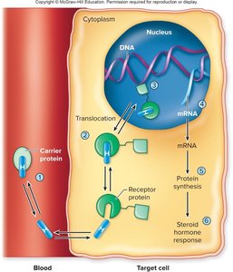
Lipophilic (fat-soluble) hormones: Receptors are located in the cytoplasm or nucleus.
Water-soluble hormones: Receptors are found on the plasma membrane.

Lipophilic Hormones and Nuclear Receptors
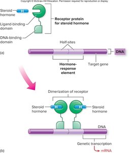
Lipophilic hormones (e.g., steroid and thyroid hormones) travel in the blood bound to carrier proteins. At the target cell, they dissociate and diffuse through the plasma membrane to bind intracellular receptors. These receptors act as transcription factors, regulating gene expression and protein synthesis.
Receptor proteins have two domains: a ligand-binding domain (for the hormone) and a DNA-binding domain (for specific DNA sequences called hormone response elements).
Hormone binding activates the DNA-binding domain, initiating transcription of target genes.
Water-Soluble Hormones and Second Messengers
Water-soluble hormones cannot cross the plasma membrane. Instead, they bind to surface receptors and activate intracellular signaling pathways using second messengers. The three main second messenger systems are:
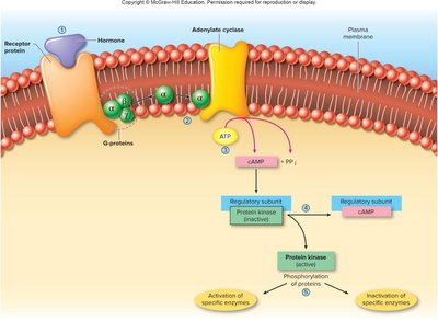
Adenylate Cyclase (cAMP) System
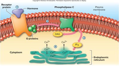
Phospholipase C System
Tyrosine Kinase System
Adenylate Cyclase (cAMP) System
Hormone binding activates adenylate cyclase via G-proteins, converting ATP to cAMP, which then activates protein kinases to elicit cellular responses.
Phospholipase C System
Hormone binding activates phospholipase C, generating IP3 and DAG, which increase intracellular Ca2+ and activate protein kinases.
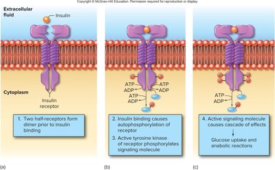
Tyrosine Kinase System
Hormone binding causes receptor dimerization and autophosphorylation, triggering a cascade of intracellular signaling events (e.g., insulin action).
The Endocrine Glands
Major Endocrine Organs
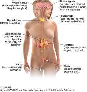
The endocrine system consists of several glands that secrete hormones directly into the bloodstream. Major glands include:
Hypothalamus
Pituitary gland
Pineal gland
Thyroid and parathyroid glands
Thymus
Adrenal glands
Pancreas
Ovaries and testes
Placenta (during pregnancy)
Hypothalamus and Pituitary Gland
Overview
The pituitary gland is often called the "master gland" because it regulates many other endocrine glands. The hypothalamus controls pituitary function via hormonal and neural signals.
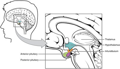
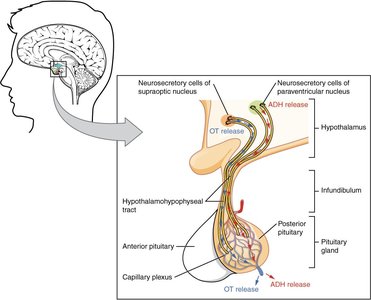
Posterior Pituitary (Neurohypophysis)
The posterior pituitary stores and releases hormones produced by the hypothalamus, specifically antidiuretic hormone (ADH) and oxytocin. These hormones are transported down axons from the hypothalamus and released into the bloodstream in response to neural signals.
ADH: Stimulates water reabsorption by the kidneys.
Oxytocin: Stimulates uterine contractions during childbirth and milk ejection during breastfeeding.
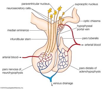
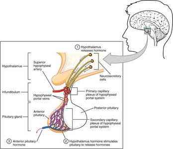
Anterior Pituitary (Adenohypophysis)
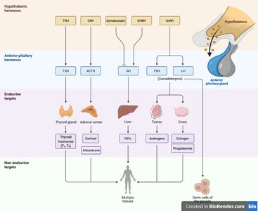
The anterior pituitary synthesizes and secretes its own hormones, but is regulated by hypothalamic hormones delivered via the hypophyseal portal system. Major anterior pituitary hormones include:
Growth hormone (GH): Stimulates growth of body tissues.
Prolactin (PRL): Promotes milk production.
Thyroid-stimulating hormone (TSH): Stimulates thyroid hormone release.
Adrenocorticotropic hormone (ACTH): Stimulates adrenal cortex hormone release.
Follicle-stimulating hormone (FSH): Stimulates gamete production.
Luteinizing hormone (LH): Stimulates androgen production.
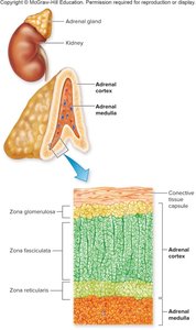
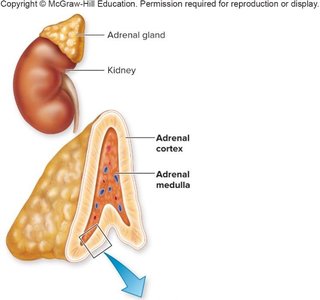
Adrenal Glands
Structure and Function
The adrenal glands are located above the kidneys and consist of two regions:
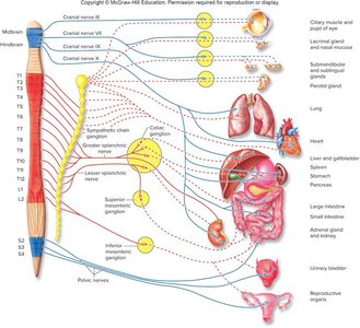
Adrenal medulla: Neuroendocrine tissue that secretes epinephrine and norepinephrine in response to sympathetic stimulation.
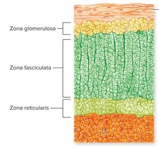
Adrenal cortex: Glandular tissue that secretes steroid hormones (corticosteroids) in response to ACTH or other signals. It has three layers: zona glomerulosa, zona fasciculata, and zona reticularis.
Adrenal Medulla
Secretes epinephrine and norepinephrine during sympathetic activation (fight-or-flight response).
Effects include increased heart rate, respiratory rate, alertness, and metabolic rate.
Adrenal Cortex
The adrenal cortex produces three main classes of steroid hormones:
Class | Layer | Main Hormone | Function |
|---|---|---|---|
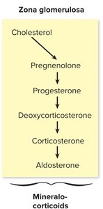
Mineralocorticoids | Zona glomerulosa | Aldosterone | Regulate Na+ and K+ balance |
Glucocorticoids | Zona fasciculata | Cortisol | Regulate metabolism, immune response, stress |
Adrenal androgens | Zona reticularis | DHEA | Supplement gonadal sex hormones |
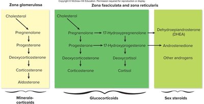
Aldosterone
Regulates blood pressure by increasing sodium reabsorption and potassium excretion in the kidneys.
Stimulated by the renin-angiotensin-aldosterone system and, to a lesser extent, by ACTH.
Deficiency leads to Addison's disease (low blood pressure, lethargy); excess causes hypertension and increased blood volume.
Cortisol
Regulated by the hypothalamic-pituitary-adrenal (HPA) axis: CRH (hypothalamus) → ACTH (anterior pituitary) → cortisol (adrenal cortex).
Functions: increases blood glucose, regulates metabolism, suppresses inflammation, affects memory, and helps the body respond to stress.
Excess cortisol (Cushing's syndrome) causes fat redistribution, muscle wasting, and other symptoms.
Stress and the Adrenal Gland
General Adaptation Syndrome (GAS)
The body's response to stress involves the adrenal gland and is described in three stages:
Alarm Reaction: Immediate activation of the adrenal glands, increased heart rate, and release of adrenaline and cortisol (fight-or-flight response).
Stage of Resistance: Body adapts to ongoing stress, cortisol levels may remain elevated.
Stage of Exhaustion: Prolonged stress leads to depletion of resources, increased risk of illness or death.
Chronic Stress and Disease
Chronic elevation of cortisol can contribute to depression, anxiety, memory problems, and insulin resistance (worsening diabetes).
Cushing's syndrome results from chronic glucocorticoid excess, causing characteristic fat redistribution and other symptoms.
