 Back
BackThe Endocrine System: Structure, Function, and Regulation
Study Guide - Smart Notes
 Tailored notes based on your materials, expanded with key definitions, examples, and context.
The Endocrine System
Overview of the Endocrine System
The endocrine system is a collection of glands and organs that secrete hormones directly into the bloodstream to regulate various physiological processes. It plays a critical role in maintaining homeostasis, growth, metabolism, and reproduction.
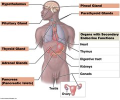
Primary endocrine organs: Hypothalamus, pituitary gland, thyroid gland, adrenal glands, pancreas (pancreatic islets), pineal gland, parathyroid glands
Organs with secondary endocrine functions: Heart, thymus, digestive tract, kidneys, gonads
Hormone Classes
Hormones are classified based on their chemical structure, which determines their mechanism of action and solubility.
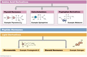
Amino acid derivatives: Include thyroid hormones, catecholamines (e.g., epinephrine), and tryptophan derivatives (e.g., melatonin)
Peptide hormones: Chains of amino acids (e.g., insulin, growth hormone)
Lipid derivatives: Include eicosanoids (e.g., prostaglandins) and steroid hormones (e.g., estrogen)
Hormone Action and Regulation
Hormone Receptors and Target Cells
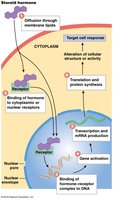
Hormones affect target cells by binding to specific receptors located in the plasma membrane, cytoplasm, or nucleus. A cell must have the appropriate receptor to respond to a hormone.
Extracellular receptors: Located on the plasma membrane; bind water-soluble hormones (e.g., peptide hormones)
Intracellular receptors: Located in the cytoplasm or nucleus; bind lipid-soluble hormones (e.g., steroid hormones)
Major Endocrine Organs and Their Functions
Hypothalamus
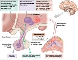
The hypothalamus integrates the nervous and endocrine systems and controls many endocrine organs through three main mechanisms:
Synthesizes antidiuretic hormone (ADH) and oxytocin (OXT), which are transported to and released by the posterior pituitary
Secretes regulatory hormones that control the anterior pituitary gland
Directly stimulates the adrenal medulla via neural pathways
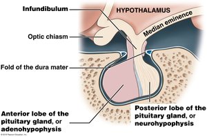
Pituitary Gland (Hypophysis)
The pituitary gland is a small, oval gland located within the sella turcica of the sphenoid bone. It releases nine peptide hormones: seven from the anterior lobe (adenohypophysis) and two from the posterior lobe (neurohypophysis).
Anterior lobe: Releases tropic hormones that regulate other endocrine glands
Posterior lobe: Releases ADH and OXT
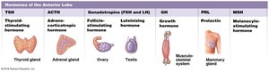
Hormones of the Anterior Lobe
Hormone | Target |
|---|---|
TSH (Thyroid-stimulating hormone) | Thyroid gland |
ACTH (Adrenocorticotropic hormone) | Adrenal gland |
FSH & LH (Gonadotropins) | Ovary, Testis |
GH (Growth hormone) | Musculoskeletal system |
PRL (Prolactin) | Mammary gland |
MSH (Melanocyte-stimulating hormone) | Melanocytes |
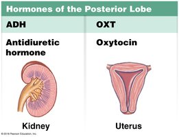
Hormones of the Posterior Lobe
Hormone | Target |
|---|---|
ADH (Antidiuretic hormone) | Kidney |
OXT (Oxytocin) | Uterus |
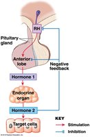
Regulation of Endocrine Activity: Negative Feedback
Negative feedback mechanisms control the activity of the hypothalamus and pituitary gland. Typically, the release of a hormone is regulated by the concentration of another hormone or the effect it produces.
Example: Hypothalamic releasing hormone triggers anterior pituitary hormone release, which stimulates a target gland to release a second hormone. The second hormone inhibits further release of the first two hormones.
Thyroid and Parathyroid Glands
Thyroid Gland
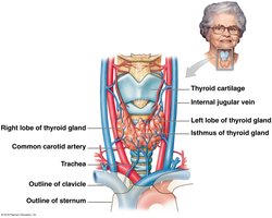
The thyroid gland is located on the anterior surface of the trachea and consists of two lobes connected by an isthmus. It requires iodine to produce thyroid hormones, which regulate metabolic rate and calcium ion levels.
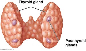
Parathyroid Glands
The parathyroid glands are two pairs of small glands embedded in the posterior surface of the thyroid gland. They secrete parathyroid hormone (PTH), the primary regulator of blood calcium levels.
Principal cells: Produce PTH, which increases calcium levels in extracellular fluids
Oxyphil cells: Function unknown
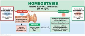
Calcium Homeostasis
PTH is released when blood calcium levels fall below normal, causing an increase in calcium levels by acting on bones, kidneys, and the digestive tract.
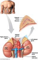
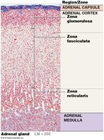
Adrenal Glands
Structure and Function
The adrenal glands, also called suprarenal glands, sit on the superior border of each kidney. They have a rich blood supply and are divided into two main regions:
Adrenal cortex: Produces corticosteroids (over 24 steroid hormones) that regulate metabolism and are vital for life
Adrenal medulla: Produces epinephrine and norepinephrine, which are involved in the sympathetic response (fight or flight)
Adrenal Medulla
The adrenal medulla secretes epinephrine and norepinephrine in response to sympathetic nervous system stimulation, resulting in increased cardiac activity, blood pressure, and blood glucose levels.
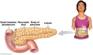
Pancreas and Glucose Regulation
Pancreatic Islets
The pancreas contains clusters of hormone-producing cells called pancreatic islets (islets of Langerhans), which make up about 1% of the organ's volume. The exocrine pancreas produces digestive enzymes, while the endocrine pancreas regulates blood glucose.
Pancreatic Islet Cells and Hormones
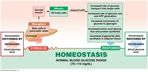
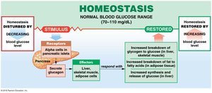
Alpha cells: Produce glucagon, which raises blood glucose by increasing glycogen breakdown and glucose release by the liver
Beta cells: Produce insulin, which lowers blood glucose by increasing glucose uptake and utilization by cells and promoting glycogen synthesis

Glucose Homeostasis
Insulin and glucagon have opposing effects to maintain blood glucose within a narrow range (70–110 mg/dL).
As blood glucose rises, beta cells secrete insulin, stimulating glucose uptake by cells
As blood glucose falls, alpha cells secrete glucagon, stimulating glycogen breakdown and glucose release
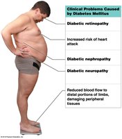
Diabetes Mellitus
Diabetes mellitus is an endocrine disorder characterized by chronic hyperglycemia (high blood glucose). It results from inadequate insulin production or abnormal insulin receptor function.
Type 1 diabetes: Inadequate insulin production; requires insulin therapy
Type 2 diabetes: Insulin resistance; associated with obesity and can often be managed with lifestyle changes and medication
Untreated diabetes disrupts metabolism, leading to tissue damage, cardiovascular disease, kidney failure, nerve degeneration, and increased risk of infection and amputation.
Endocrine Disorders
Causes and Effects
Endocrine disorders can result from abnormalities at the gland, regulatory mechanisms, or target tissues. Most disorders are due to hypo-secretion (underproduction) or hyper-secretion (overproduction) of hormones. Abnormal receptor function can also cause endocrine disease, as seen in type 2 diabetes.
