Back
BackThe Endocrine System: Structure, Function, and Hormonal Regulation
Study Guide - Smart Notes
 Tailored notes based on your materials, expanded with key definitions, examples, and context.
The Endocrine System
Overview and Historical Context
The endocrine system is a major regulatory system in the human body, responsible for producing hormones that control various physiological processes. Hormones were first proposed as regulatory molecules in 1902, and many diseases are linked to hormone dysfunction. External substances, such as alcohol, can affect endocrine organs like the testes and parathyroid glands.
Functions of the Endocrine System
Hormone Production: Endocrine glands produce hormones that regulate body functions.
Transport: Hormones travel through the bloodstream to target tissues.
Duration: Hormonal effects are typically longer-lasting than those of the nervous system.
Endocrine Glands and Organs
Endocrine glands: Ductless glands whose products enter lymphatic ducts or blood vessels.
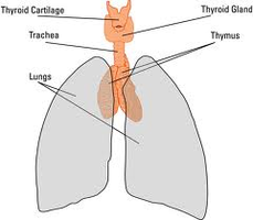
Pure endocrine organs: Pituitary, thyroid, parathyroid, pineal, adrenal gland, and thymus.
Neuroendocrine organ: Hypothalamus.
Mixed-function organs: Pancreas, ovaries, placenta, testes.
Other tissues: Stomach, kidneys, small intestines, heart, and most other tissues contain pockets of endocrine cells.
Hormonal Effects and Target Tissues
Cellular Changes: Hormones can alter membrane permeability, stimulate protein synthesis, activate/deactivate enzymes, induce secretory activity, and stimulate mitosis.
Target Tissue Specificity: Some hormones act on specific tissues (e.g., TSH activates only thyroid cells), while others have broad effects (e.g., thyroxine affects nearly every cell).
Target Cell Activation
Activation depends on:
Amount of hormone in the blood
Number of receptors in the cell
Strength of the bond between hormone and receptor
Increasing any of these factors results in a stronger effect. Receptor numbers can vary based on physiological conditions.
Hormone Activity
Hormones are effective in small quantities.
Duration in circulation varies widely.
Onset of effects can be immediate or delayed.
Duration of effects ranges from seconds to hours.
Control of Hormone Release
Humoral stimuli: Changes in blood ions or nutrients trigger hormone release.
Neural stimuli: Nerve fibers stimulate hormone release.
Hormonal stimuli: Presence of one hormone triggers release of another.
Examples of Stimuli
Humoral: Low Ca2+ triggers parathyroid hormone; high blood sugar triggers insulin.
Neural: Sympathetic system triggers adrenal cortex to release catecholamines.
Hormonal: TSH stimulates thyroid to release thyroxine.
Pituitary Gland (Hypophysis)
Structure and Function
The pituitary gland secretes at least eight hormones and is attached to the hypothalamus by the infundibulum. It consists of two lobes: the posterior pituitary and the anterior pituitary.
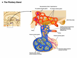
Posterior Pituitary (Neurohypophysis)
Composed of neural tissue; part of the brain.
Stores and releases hormones made by the hypothalamus: Oxytocin and Antidiuretic hormone (ADH).
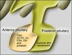
Oxytocin
Causes uterine contractions.
Involved in orgasm and milk let-down.
Promotes nurturing and affectionate behavior; bonding in new parents and relationships.
Antidiuretic Hormone (ADH)
Release is triggered by concentrated blood.
Causes kidneys to reabsorb more water and produce concentrated urine.
Anterior Pituitary (Adenohypophysis)
Connected to the hypothalamus by a portal system (two capillary beds connected by venules).
Hypothalamus can stimulate or inhibit anterior pituitary.
Makes and secretes six hormones:
Growth Hormone (GH)
Prolactin
Thyroid Stimulating Hormone (TSH)
Adrenocorticotropic Hormone (ACTH)
Follicle Stimulating Hormone (FSH)
Luteinizing Hormone (LH)
Growth Hormone (GH)
Stimulated by Growth Hormone Releasing Hormone (GHRH) from hypothalamus.
Encourages fat use for energy, conserves glucose, stimulates protein production.
Inhibited by Growth Hormone Inhibiting Hormone (GHIH).
Prolactin
Inhibited by prolactin-inhibiting hormone (PIH)/dopamine.
Absence of PIH allows prolactin-releasing hormone secretion.
Stimulates milk production.
Thyroid Stimulating Hormone (TSH)
Stimulated by thyrotropin-releasing hormone (TRH) from hypothalamus.
Causes thyroid to release hormones.
Inhibited by presence of thyroid hormones.
Adrenocorticotropic Hormone (ACTH)
Stimulated by corticotropic-releasing hormone from hypothalamus.
Stimulates adrenal cortex to release glucocorticoids and androgens.
Inhibited by presence of glucocorticoids.
Gonadotropins (FSH and LH)
Production begins at puberty, stimulated by Gonadotropin Releasing Hormone (GnRH).
Inhibited by feedback inhibition.
FSH stimulates gamete production; LH matures follicle in females and stimulates testosterone release in males.
Thyroid Gland
Structure and Function
The thyroid gland is located on the trachea, inferior to the larynx, and is the largest pure endocrine gland. Follicle cells produce thyroid hormone, while parafollicular cells produce calcitonin.
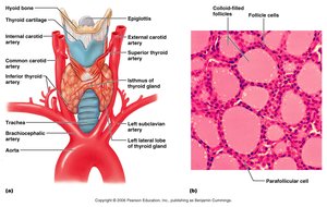
Thyroid Hormones
Two hormones: Thyroxine (T4) and Triiodothyronine (T3).
Stimulate nearly every cell in the body.
Regulate basal metabolic rate (BMR), growth, and blood pressure.
Dysfunction can cause hypothyroidism or hyperthyroidism.
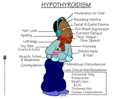
Hypothyroidism
Low metabolic rate, intolerance to cold, puffy eyes, lethargy, weight gain.
Can be caused by lack of iodine.
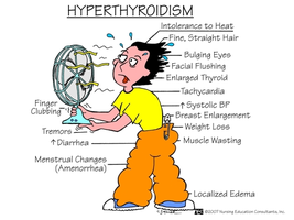
Hyperthyroidism (Grave's Disease)
High metabolic rate, sweating, bulging eyes, nervousness, weight loss.
Calcitonin
Pharmacological doses lower blood Ca2+ by inhibiting osteoclasts and stimulating Ca2+ deposition in bone.
Parathyroid Gland
Structure and Function
Usually four glands located on the posterior side of the thyroid.
Chief cells secrete parathyroid hormone (PTH).
Parathyroid Hormone (PTH)
Release stimulated by low blood Ca2+.
Stimulates osteoclasts to break down bone.
Enhances Ca2+ reabsorption in kidneys.
Increases Ca2+ absorption in intestines by activating vitamin D.
Adrenal Glands
Structure and Function
Adrenal medulla: part of the sympathetic nervous system.
Adrenal cortex: produces over two dozen steroid hormones (corticosteroids).
Adrenal Medulla
Chromaffin cells produce catecholamines (epinephrine and norepinephrine).
Causes short-term sympathetic response throughout the body.
Adrenal Cortex
Three layers:
Zona glomerulosa: Produces mineralocorticoids (e.g., aldosterone) to regulate electrolyte concentration, especially Na+ and K+.
Zona fasciculata: Produces glucocorticoids (e.g., cortisol) to influence metabolism and resist stress.
Zona reticularis: Produces gonadocorticoids (weak androgens), which are converted to testosterone and estrogen.
Pancreas
Structure and Function
Located behind the stomach; both endocrine and exocrine functions.
Pancreatic islets (islets of Langerhans) contain alpha and beta cells.
Alpha Cells (α)
Produce glucagon, which stimulates the liver to break down glycogen into glucose, raising blood sugar levels.
Beta Cells (β)
Produce insulin, which stimulates tissues to take up sugar and the liver to make glycogen from glucose.
Diabetes Mellitus
Type 1: Not enough insulin is made.
Type 2: Insulin is made but not functioning properly.
Sugar in diet cannot be absorbed; symptoms include sugar and ketones in urine, weight loss, excessive urine production, thirst, and hunger.
Pineal Gland
Structure and Function
Located in the brain; produces melatonin.
Receives input from eyes to regulate daily sleep/wake cycles.
Sunlight suppresses melatonin production.
Thymus
Structure and Function
Located deep to the sternum; large in children.
Produces hormones needed for normal T lymphocyte development.