Back
The Endocrine System: Structure, Function, and Hormonal Regulation
Study Guide - Smart Notes
 Tailored notes based on your materials, expanded with key definitions, examples, and context.
The Endocrine System
Overview and Function
The endocrine system is a network of glands that produce hormones to regulate various physiological processes throughout the body. Unlike the nervous system, which acts quickly and locally, endocrine signals are slower and longer-lasting, affecting distant target tissues via the bloodstream.
Hormones: Chemical messengers first proposed in 1902; dysfunction can cause many diseases.
Endocrine glands: Ductless glands whose products enter blood or lymphatic vessels.
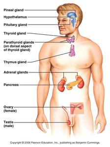
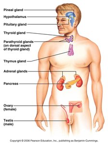
Endocrine organs: Scattered throughout the body, including pure endocrine organs (pituitary, thyroid, parathyroid, pineal, adrenal, thymus), neuroendocrine (hypothalamus), and organs with endocrine tissue (pancreas, ovaries, placenta, testes).
Hormonal Effects and Target Tissue
Hormones can cause a variety of changes in target tissues, which may be highly specific or broadly distributed.
Effects: Alter membrane permeability, stimulate protein synthesis, activate/deactivate enzymes, induce secretory activity, and stimulate mitosis.
Target tissue: The specific tissue where a hormone exerts its effect (e.g., TSH acts only on thyroid cells; thyroxine affects nearly all cells).
Activation: Depends on hormone concentration, receptor number, and bond strength between hormone and receptor.
Control of Hormone Release
Hormone release is regulated by three main stimuli:
Humoral stimuli: Changes in blood ion/nutrient levels (e.g., low Ca2+ triggers parathyroid hormone).
Neural stimuli: Nerve fibers stimulate hormone release (e.g., sympathetic system triggers adrenal medulla).
Hormonal stimuli: One hormone triggers release of another (e.g., TSH stimulates thyroxine release).
Pituitary Gland (Hypophysis)
Structure and Function
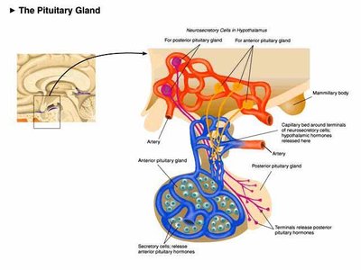
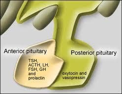
The pituitary gland is a central endocrine organ attached to the hypothalamus by the infundibulum. It consists of two lobes:
Posterior pituitary (neurohypophysis): Neural tissue; stores and releases hormones made by the hypothalamus (oxytocin and ADH).
Anterior pituitary (adenohypophysis): Glandular tissue; makes and secretes six hormones.
Posterior Pituitary Hormones
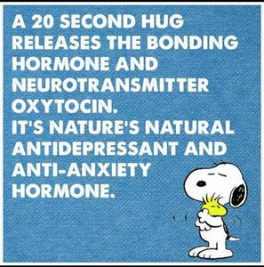
Oxytocin: Causes uterine contractions, milk let-down, and promotes bonding and affectionate behavior.
Antidiuretic hormone (ADH): Triggered by concentrated blood; causes kidneys to reabsorb water and produce concentrated urine.
Anterior Pituitary Hormones
Growth Hormone (GH): Stimulated by GHRH; promotes fat use, protein synthesis, and growth. Inhibited by GHIH.
Prolactin: Stimulates milk production; regulated by PIH (dopamine).
Tropins/Trophic hormones:
Thyroid Stimulating Hormone (TSH): Stimulates thyroid hormone release.
Adrenocorticotropic hormone (ACTH): Stimulates adrenal cortex to release glucocorticoids and androgens.
Follicle Stimulating Hormone (FSH): Stimulates gamete production.
Luteinizing Hormone (LH): Matures follicle in females, stimulates testosterone in males.

Thyroid Gland
Structure and Function
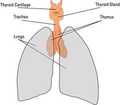
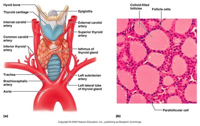
The thyroid gland is located on the trachea, inferior to the larynx, and is the largest pure endocrine gland.
Follicle cells: Produce thyroid hormone (T3 and T4).
Parafollicular cells: Produce calcitonin.
Thyroid Hormone
Thyroxine (T4) and Triiodothyronine (T3): Stimulate nearly every cell, regulate basal metabolic rate (BMR), growth, and blood pressure.
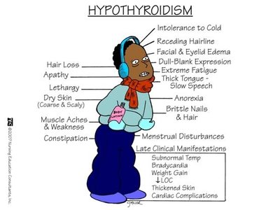
Hypothyroidism: Low metabolic rate, intolerance to cold, lethargy, weight gain; often caused by iodine deficiency.
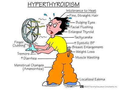
Hyperthyroidism: High metabolic rate, sweating, bulging eyes, nervousness, weight loss; also known as Grave's Disease.
Calcitonin
Pharmacological doses: Lowers blood Ca2+ by inhibiting osteoclasts and stimulating Ca2+ deposition in bone.
Parathyroid Gland
Structure and Function
Usually four glands located on the posterior side of the thyroid. Chief cells secrete parathyroid hormone (PTH). 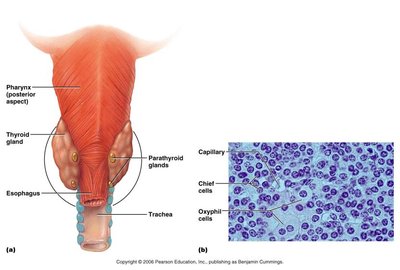
Parathyroid Hormone (PTH)
Release stimulated by low blood Ca2+.
Stimulates osteoclasts to break down bone.
Enhances Ca2+ reabsorption in kidneys.
Increases Ca2+ absorption in intestines by activating vitamin D.
Adrenal Glands
Structure and Function
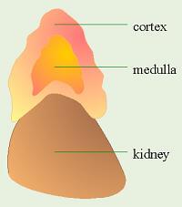
The adrenal glands are located atop the kidneys and consist of two main regions:
Adrenal medulla: Part of the sympathetic nervous system; chromaffin cells produce catecholamines (epinephrine and norepinephrine).
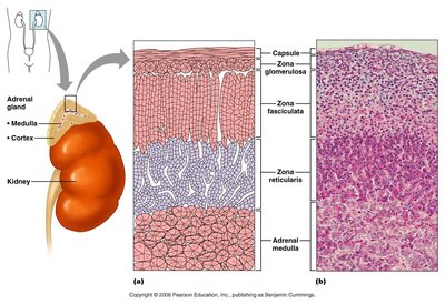
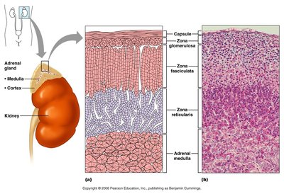
Adrenal cortex: Produces over two dozen steroid hormones (corticosteroids) in three layers.
Adrenal Cortex Layers
Zona glomerulosa: Most superficial; produces mineralocorticoids (e.g., aldosterone) to regulate Na+ and K+ balance.
Zona fasciculata: Middle layer; produces glucocorticoids (e.g., cortisol) to influence metabolism and resist stress.
Zona reticularis: Innermost layer; produces gonadocorticoids (weak androgens), contributing to puberty and female sex drive.
Pancreas
Structure and Function
The pancreas is located behind the stomach and has both endocrine and exocrine functions.
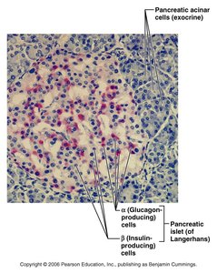
Pancreatic islets (Islets of Langerhans): Contain alpha and beta cells.
Alpha cells (α): Produce glucagon, which raises blood sugar by stimulating glycogen breakdown in the liver.
Beta cells (β): Produce insulin, which lowers blood sugar by stimulating tissue uptake and glycogen formation.
Diabetes Mellitus
Type 1: Insufficient insulin production.
Type 2: Insulin produced but not functioning properly.
Symptoms: Sugar and ketones in urine, excessive urination, thirst, hunger, and weight loss.
Pineal Gland
Structure and Function
The pineal gland is located in the brain and produces melatonin, which regulates daily sleep/wake cycles. Sunlight suppresses melatonin production. 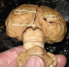
Thymus
Structure and Function
The thymus is located deep to the sternum and is large in children. It produces hormones necessary for normal T lymphocyte development, which are essential for immune function.