Back
BackThe Endocrine System: Structure, Function, and Regulation
Study Guide - Smart Notes
 Tailored notes based on your materials, expanded with key definitions, examples, and context.
The Endocrine System
Overview of the Endocrine System
The endocrine system is one of the two major regulatory systems of the body, alongside the nervous system. It consists of organs that synthesize and secrete chemical messengers called hormones into the bloodstream. Hormones interact with specific cells known as target cells, which possess receptors for these hormones, leading to changes in cellular function. The tissues containing these target cells are referred to as target tissues.
Hormones: Chemical messengers secreted by endocrine glands.
Target Cells: Cells with specific receptors for hormones.
Receptors: Proteins that bind hormones and initiate cellular responses.
Comparison of the Endocrine and Nervous Systems
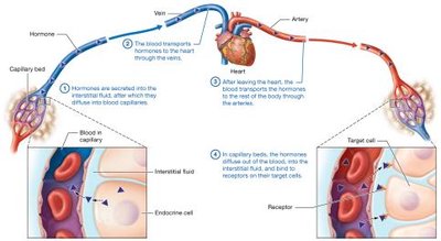
The endocrine system regulates physiological processes by releasing hormones into the blood, which travel to distant target cells. In contrast, the nervous system uses direct synaptic connections for rapid communication.
Endocrine cells secrete hormones into interstitial fluid, which then diffuse into blood capillaries.
Hormones are transported by the blood to the heart and then distributed throughout the body.
Hormones diffuse out of capillaries and bind to receptors on target cells.
Effects of hormones are generally slower but longer-lasting than nervous system signals.
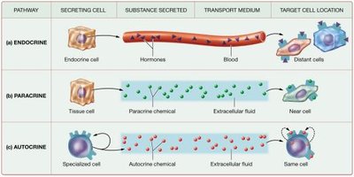
Types of Signaling: Endocrine, Paracrine, and Autocrine
Hormones can act through different signaling pathways:
Endocrine: Hormones are secreted into the blood and affect distant cells.
Paracrine: Chemicals are secreted into the extracellular fluid to influence nearby cells.
Autocrine: Chemicals are secreted by a cell to affect itself.
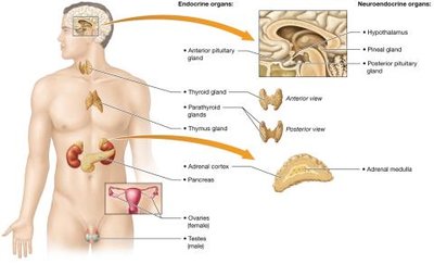
Overview of Endocrine Organs
Endocrine glands are ductless organs that secrete hormones directly into the interstitial fluid for transport by the bloodstream. Primary endocrine organs include:
Anterior pituitary gland
Thyroid gland
Parathyroid glands
Adrenal cortices
Endocrine pancreas
Thymus
Ovaries or testes
Secondary endocrine glands (e.g., heart, kidneys, small intestine, adipose tissue) produce hormones but belong to other systems. Neuroendocrine organs (hypothalamus, pineal gland, adrenal medulla) consist of nervous tissue but secrete neurohormones.
Hormones: Structure, Transport, and Action
Classes of Hormones
Hormones are classified based on their chemical structure:
Amino Acid-Based Hormones: Derived from amino acids; mostly hydrophilic.
Peptide/Protein Hormones: Chains of amino acids; generally hydrophilic.
Steroid Hormones: Derived from cholesterol; hydrophobic and lipid-soluble.
Hormone Transport in Blood
Free Hormones: Small, hydrophilic hormones travel freely in plasma.
Bound Hormones: Hydrophobic hormones form complexes with binding proteins, allowing transport and extending their lifespan in blood.
Target Cells and Receptors
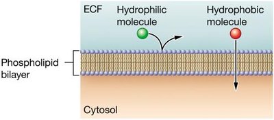
Hormones bind to specific receptors on or within target cells. Some hormones bind to a single receptor type, while others bind to multiple types, producing different effects. Receptors may be located:
In the plasma membrane (for hydrophilic hormones)
In the cytosol or nucleus (for hydrophobic hormones)
Mechanisms of Hormone Action
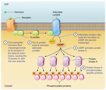
Hydrophilic hormones typically act via second-messenger systems, such as the adenylate cyclase–cAMP pathway:
Hormone binds to receptor, activating G-protein.
G-protein activates adenylate cyclase, which forms cAMP from ATP.
cAMP activates protein kinase A, which phosphorylates proteins, altering their activity.
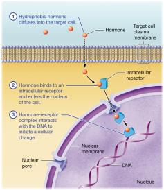
Hydrophobic hormones diffuse into target cells, bind to intracellular receptors, and directly influence gene expression by interacting with DNA.
Regulation of Hormone Secretion
Types of Stimuli for Hormone Secretion
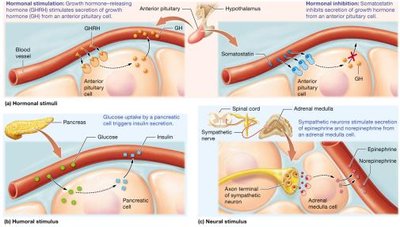
Hormone secretion is initiated by:
Hormonal Stimuli: Response to other hormones.
Humoral Stimuli: Response to changes in ion or compound concentration in blood.
Neural Stimuli: Response to nervous system signals.
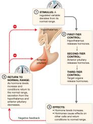
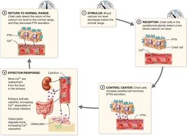
Negative Feedback Regulation
Hormone secretion is typically regulated by negative feedback loops:
Stimulus: Physiological variable deviates from normal range.
Receptor: Endocrine cell receptors detect deviation.
Control Center: Endocrine cell adjusts hormone secretion.
Effector/Response: Hormone triggers response to restore normal range.
Return to Normal Range: Secretion returns to baseline.

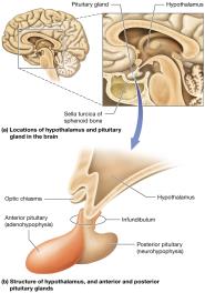
Hypothalamus and Pituitary Gland
Structure and Functional Relationships
The hypothalamus connects to the pituitary gland via the infundibulum. The anterior pituitary (adenohypophysis) is a true gland, while the posterior pituitary (neurohypophysis) is nervous tissue that stores neurohormones.
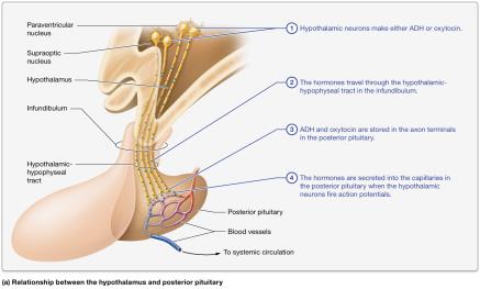
Hormones of the Hypothalamus and Posterior Pituitary
The posterior pituitary stores and releases antidiuretic hormone (ADH) and oxytocin:
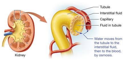
ADH: Promotes water retention by kidneys; decreases urine production.
Oxytocin: Stimulates uterine contractions and milk ejection; operates via positive feedback.
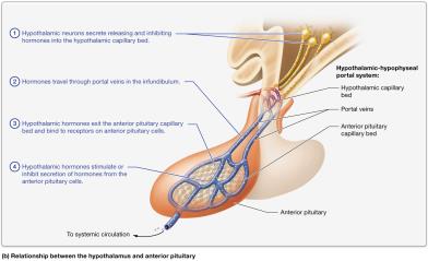
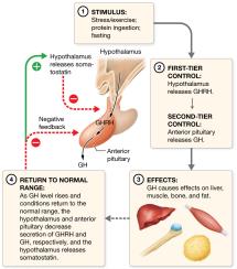
Functional Relationship of the Hypothalamus and Anterior Pituitary
The hypothalamus produces releasing and inhibiting hormones that control the anterior pituitary. Tropic hormones from the anterior pituitary regulate other endocrine glands.
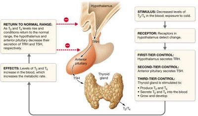
Hormone secretion is regulated by multi-tiered negative feedback loops:
First tier: Hypothalamic neuroendocrine cells
Second tier: Anterior pituitary cells
Third tier: Target tissue cells
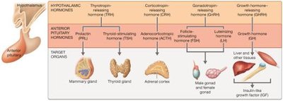
Anterior Pituitary Hormones
Anterior pituitary hormones include:
TSH: Stimulates thyroid gland
ACTH: Stimulates adrenal cortex
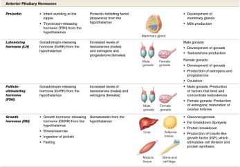
Prolactin: Stimulates mammary glands
LH: Stimulates gonads
FSH: Stimulates gonads
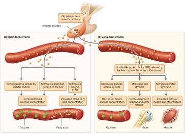
GH: Stimulates growth and metabolism
Hormones of the Hypothalamus and Pituitary Gland (Table)
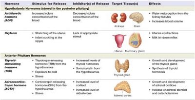
Hormone | Stimulus for Release | Inhibitor of Release | Target Tissue(s) | Effects |
|---|---|---|---|---|
Antidiuretic hormone (ADH) | Increased solute concentration of blood | Decreased solute concentration of blood | Kidneys, brain | Water reabsorption, increased blood volume |
Oxytocin | Stretching of uterus, infant suckling | Lack of appropriate stimulus | Uterus, mammary gland | Uterine contraction, milk let-down reflex |
TSH | TRH from hypothalamus, exposure to cold | Somatostatin from hypothalamus, increased levels of thyroid hormones | Thyroid gland | Growth and development, secretion of thyroid hormones |
ACTH | CRH from hypothalamus, stress | Increased levels of cortisol and aldosterone | Adrenal cortex | Growth and development, release of adrenal corticosteroids |
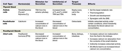
Thyroid and Parathyroid Glands
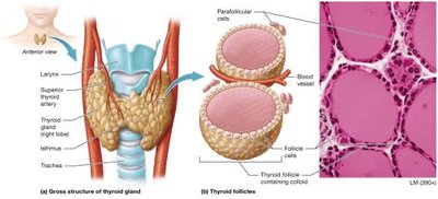
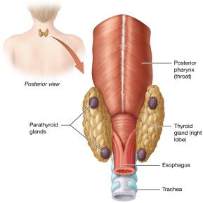
Structure of the Thyroid and Parathyroid Glands
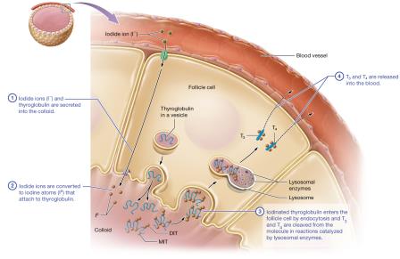
The thyroid gland is located in the anterior neck and consists of two lobes connected by the isthmus. It contains thyroid follicles, follicle cells, colloid, and parafollicular cells. The parathyroid glands are usually four small glands embedded in the posterior thyroid, with chief cells producing parathyroid hormone (PTH).
Thyroid Hormones: Metabolic Regulators
Thyroid hormones (T3 and T4) regulate metabolic rate, thermoregulation, growth, and development. T3 is more active than T4, which is converted to T3 in target cells. These hormones are hydrophobic and act via intracellular receptors.
Increase basal metabolic rate
Stimulate ATP-requiring Na+/K+ pumps
Trigger gluconeogenesis in the liver
Promote protein and fat breakdown
Synergize with the sympathetic nervous system
Thyroid Disorders
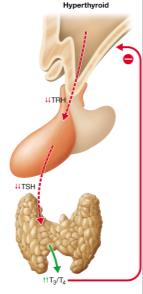
Hyperthyroidism: Overproduction of thyroid hormones (e.g., Graves disease)
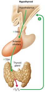
Hypothyroidism: Underproduction of thyroid hormones (e.g., Hashimoto thyroiditis, iodine deficiency)
Goiter: Enlargement of the thyroid gland due to abnormal TSH-like proteins
Congenital Hypothyroidism: Inadequate thyroid function in infants, leading to developmental delays

Parathyroid Hormone and Calcitonin: Bone Homeostasis
PTH is secreted in response to low blood calcium and increases calcium release from bone, absorption in the intestine, and reabsorption in the kidneys. Calcitonin is secreted in response to high blood calcium and inhibits osteoclast activity.
Cell Type | Hormone(s) | Stimulus for Secretion | Inhibitor(s) of Secretion | Target Tissue(s) | Effects |
|---|---|---|---|---|---|
Follicle cells (Thyroid) | T3, T4 | TSH from anterior pituitary | Increased T3/T4 | Nearly every cell | Set metabolic rate, thermoregulation, growth, SNS synergy |
Parafollicular cells (Thyroid) | Calcitonin | Increased blood calcium | Decreased blood calcium | Osteoclasts | Inhibits osteoclast activity |
Chief cells (Parathyroid) | PTH | Decreased blood calcium | Increased blood calcium | Bone, kidneys, intestines | Increase calcium reabsorption and absorption |