 Back
BackChapter 15 Objectives
Study Guide - Smart Notes
 Tailored notes based on your materials, expanded with key definitions, examples, and context.
Special Senses: The Eye and Vision: Chapter 15
Accessory Eye Structures
The accessory structures of the eye protect and support the function of the eyeball. They include the eyelids, conjunctiva, and the lacrimal apparatus, which are essential for maintaining eye health and proper vision.
Palpebrae (Eyelids): Protect the eye from foreign objects and help spread tears.
Conjunctiva: A thin, transparent mucous membrane lining the eyelids and covering the anterior surface of the eyeball, except the cornea.
Lacrimal Apparatus: Produces and drains tears, keeping the eye moist and free of debris.
Lacrimal gland: Produces tears.
Lacrimal puncta, canaliculi, sac, and nasolacrimal duct: Drain tears from the eye into the nasal cavity.
Extrinsic Eye Muscles
Six extrinsic muscles control the movement of the eyeball, allowing for precise and coordinated eye movements.
Superior, Inferior, Medial, and Lateral Rectus: Move the eye up, down, medially, and laterally, respectively.
Superior and Inferior Oblique: Rotate the eye and assist in complex movements.
Trochlea: A pulley-like structure for the superior oblique muscle.
Structure of the Eyeball
The eyeball consists of three main layers (tunics) and several internal structures that contribute to vision.
Fibrous Tunic: Outermost layer; includes the cornea (transparent, refracts light) and sclera (white, protective).
Vascular Tunic (Uvea): Middle layer; includes the iris (controls pupil size), ciliary body (produces aqueous humor, controls lens shape), pupil (light entry), choroid (vascular, nourishes retina), and suspensory ligaments (hold lens).
Sensory Tunic (Neural Layer): Innermost layer; the retina contains photoreceptors and neurons for visual processing.
Internal Structures of the Eyeball
Anterior Cavity: Contains aqueous humor (nourishes cornea and lens), divided into anterior and posterior chambers by the iris.
Lens: Focuses light onto the retina.
Posterior Cavity: Contains vitreous humor (maintains eyeball shape and holds retina in place).
Eye Histology
Microscopic examination of the eye reveals specialized layers and cell types essential for vision.
Choroid: Pigmented, vascular layer that supplies nutrients to the retina.
Retina: Composed of two main layers:
Pigmented layer: Absorbs stray light and prevents reflection.
Neural layer: Contains photoreceptors (rods and cones), bipolar cells, and ganglion cells.
Photoreceptor Cells: Rods (dim light, black and white vision) and cones (color vision, sharpness).
Bipolar Cells: Transmit signals from photoreceptors to ganglion cells.
Ganglion Cells: Their axons form the optic nerve.
Key Structures Identified in Dissection
Sclera: Tough, white outer layer.
Cornea: Transparent anterior part (may appear cloudy in preserved specimens).
Optic Nerve: Transmits visual information to the brain.
Lens, Ciliary Body, Suspensory Ligaments, Iris: Involved in focusing and regulating light entry.
Vitreous Humor: Gel-like substance in the posterior cavity.
Retina: Yellowish, delicate layer; contains the optic disc (blind spot) and macula lutea/fovea centralis (area of sharpest vision).
Choroid and Tapetum Lucidum: The latter is an iridescent layer in some animals for improved night vision.
Visual Tests
Blind Spot Demonstration
The blind spot is the region of the retina (optic disc) where the optic nerve exits the eye; it contains no photoreceptors, so light focused here is not detected.
Test: By focusing on a fixed point and moving an image, the blind spot can be detected when a mark disappears from view.
Reason: The absence of rods and cones at the optic disc means no image is formed in this region.
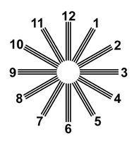
Astigmatism Test
Astigmatism is a common vision condition caused by irregular curvature of the cornea or lens, resulting in blurred or distorted vision.
Test: Viewing a chart with radiating lines (as shown below) can reveal astigmatism if some lines appear blurred or lighter than others.
Interpretation: If all lines are equally sharp, astigmatism is unlikely; if some are blurred, astigmatism may be present.
Taste: Papillae of the Tongue
The tongue contains specialized structures called papillae, which house taste buds and contribute to the sense of taste.
Fungiform Papillae: Mushroom-shaped, scattered across the tongue, contain taste buds.
Vallate Papillae: Large, arranged in a V-shape at the back of the tongue, contain many taste buds.
Foliate Papillae: Located on the sides of the tongue, contain taste buds (more prominent in children).
