 Back
The Heart: Electrical Conduction, Cardiac Action Potentials, Electrocardiogram, and Cardiac Cycle
Study Guide - Smart Notes
 Tailored notes based on your materials, expanded with key definitions, examples, and context.
The Heart
Electrical Conduction System of the Heart
The heart's intrinsic conduction system ensures coordinated and rhythmic contractions by generating and conducting action potentials. This system is essential for maintaining effective blood circulation throughout the body.
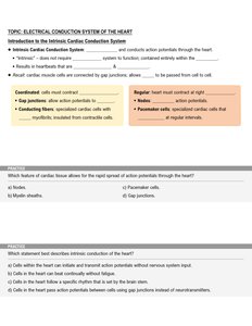
Intrinsic Cardiac Conduction System: Specialized cardiac muscle cells (pacemaker cells) generate and conduct action potentials independently of nervous system input.
Autorhythmicity: The heart can initiate its own electrical impulses due to pacemaker cells.
Gap Junctions: Cardiac muscle cells are connected by gap junctions, allowing rapid spread of action potentials.
Types of Cardiac Cells:
Contractile cells: Make up the majority of the heart muscle and are responsible for contraction.
Pacemaker cells: Initiate and conduct electrical impulses.
Anatomy of the Intrinsic Conduction System
The conduction system consists of specialized structures that generate and distribute electrical signals throughout the heart.
Sinoatrial (SA) Node: Located in the right atrium; initiates the heartbeat (the "pacemaker").
Atrioventricular (AV) Node: Located at the junction of the atria and ventricles; delays the impulse to allow atrial contraction before ventricular contraction.
Bundle of His (AV Bundle): Conducts impulses from the AV node to the ventricles.
Right and Left Bundle Branches: Carry impulses through the interventricular septum.
Purkinje Fibers: Distribute the impulse throughout the ventricles, causing contraction.
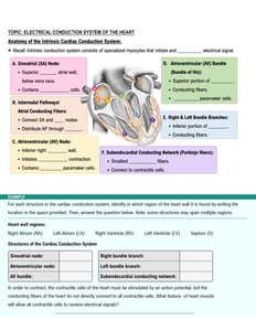
Table: Structures of the Cardiac Conduction System
Structure | Location | Function |
|---|---|---|
SA Node | Right atrium | Initiates action potential |
AV Node | Interatrial septum | Delays impulse |
Bundle of His | Interventricular septum | Conducts impulse to ventricles |
Bundle Branches | Right and left sides of septum | Carry impulse to apex |
Purkinje Fibers | Ventricular walls | Stimulate ventricular contraction |
Conduction Pathway and Steps of Cardiac Conduction
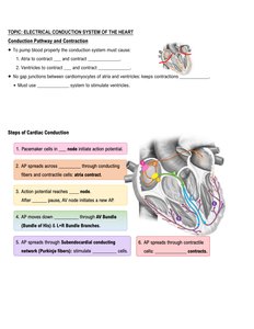
For effective pumping, the heart's conduction system must ensure that atria contract before ventricles and that both sides contract in a coordinated manner.
Pacemaker cells in the SA node initiate the action potential.
Action potential spreads through atria via internodal pathways, causing atrial contraction.
Impulse reaches the AV node, where it is delayed.
Impulse travels down the Bundle of His and bundle branches.
Impulse spreads through Purkinje fibers, causing ventricular contraction.
Control of Heart Rate
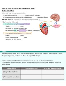
Heart rate is regulated by intrinsic pacemaker activity and extrinsic factors such as the autonomic nervous system and hormones.
Sympathetic stimulation: Increases heart rate and contractility.
Parasympathetic stimulation: Decreases heart rate (primarily via the vagus nerve).
Medullary centers: Cardioacceleratory and cardioinhibitory centers in the medulla oblongata regulate autonomic input to the heart.
Cardiac Action Potentials
Introduction to Action Potentials in Cardiac Cells
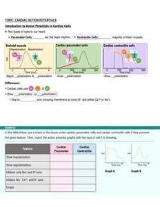
Cardiac muscle cells generate action potentials that trigger contraction. There are two main types of cardiac cells: pacemaker cells and contractile cells.
Pacemaker cells: Set the heart rhythm; have spontaneous depolarization (autorhythmicity).
Contractile cells: Make up the majority of the myocardium; responsible for contraction.
Pacemaker Cell Action Potentials
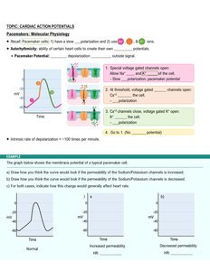
Pacemaker cells have a unique action potential characterized by a slow depolarization (pacemaker potential), rapid depolarization, and repolarization.
Phases:
Slow depolarization (due to Na+ influx)
Rapid depolarization (due to Ca2+ influx)
Repolarization (due to K+ efflux)
Autorhythmicity: Pacemaker cells can spontaneously generate action potentials.
Contractile Cell Action Potentials
Contractile cells have a prolonged action potential due to a plateau phase, which prevents tetanus and allows for effective pumping.
Phases:
Rapid depolarization (Na+ influx)
Plateau phase (Ca2+ influx balances K+ efflux)
Repolarization (K+ efflux)
Absolute refractory period: Prevents another action potential during contraction, ensuring rhythmic heartbeats.
Comparing Action Potentials in Pacemaker and Contractile Cells
Pacemaker and contractile cells differ in their ion channel activity and action potential shape.
Pacemaker cells: Slow depolarization, no true resting potential, spontaneous activity.
Contractile cells: Rapid depolarization, plateau phase, stable resting potential.
Electrocardiogram (ECG)
Electrocardiogram Basics
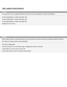
An electrocardiogram (ECG or EKG) records the electrical activity of the heart using electrodes placed on the body. It is used to assess heart rhythm and detect abnormalities.
P wave: Atrial depolarization
QRS complex: Ventricular depolarization (and atrial repolarization)
T wave: Ventricular repolarization
ECG Pathologies
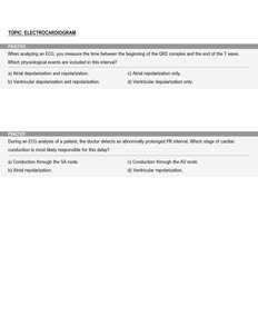
Abnormal ECG patterns can indicate arrhythmias, heart blocks, tachycardia, bradycardia, and fibrillation.
Bradycardia: Slow heart rate
Tachycardia: Fast heart rate
Fibrillation: Uncoordinated contraction; can be life-threatening if in the ventricles
Heart block: Impaired conduction between atria and ventricles
Cardiac Cycle
Systole, Diastole, Pressure, and Valves
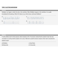
The cardiac cycle consists of alternating periods of contraction (systole) and relaxation (diastole) of the atria and ventricles. Changes in pressure open and close heart valves, ensuring unidirectional blood flow.
Systole: Contraction phase; blood is ejected from chambers.
Diastole: Relaxation phase; chambers fill with blood.
Valves: Atrioventricular (AV) valves and semilunar (SL) valves open and close in response to pressure changes.
Events in the Cardiac Cycle
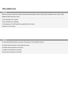
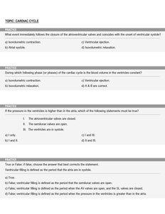
The cardiac cycle is divided into phases based on whether the ventricles are in systole or diastole and whether blood is entering or leaving the chambers.
Ventricular filling: Blood flows from atria to ventricles (diastole).
Isovolumetric contraction: Ventricles contract with all valves closed (systole).
Ventricular ejection: Blood is pumped into arteries (systole).
Isovolumetric relaxation: Ventricles relax with all valves closed (diastole).
Heart Sounds
Heart sounds are produced by the closing of heart valves. The first sound (S1) is due to AV valve closure; the second sound (S2) is due to semilunar valve closure. Abnormal sounds (murmurs) may indicate valve pathology.
S1 ("lub"): Closure of AV valves at the start of ventricular systole.
S2 ("dub"): Closure of semilunar valves at the start of ventricular diastole.
Cardiac Cycle: Integrating All Events
All events of the cardiac cycle can be visualized in a single diagram showing pressure, volume, and valve status over time. This integration helps in understanding the timing of heart sounds, valve movements, and blood flow.
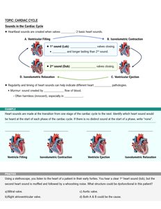
Table: Valve Status During Cardiac Cycle
Phase | AV Valves | SL Valves |
|---|---|---|
Ventricular Filling | Open | Closed |
Isovolumetric Contraction | Closed | Closed |
Ventricular Ejection | Closed | Open |
Isovolumetric Relaxation | Closed | Closed |
Additional info: The notes above are expanded with academic context to ensure clarity and completeness for ANP college students. All images included are directly relevant to the adjacent explanations and reinforce the educational content.
