 Back
BackThe Heart: Structure, Blood Flow, and Valves
Study Guide - Smart Notes
 Tailored notes based on your materials, expanded with key definitions, examples, and context.
The Heart and Circulatory Pathways
Overview of Heart Function
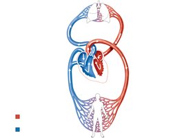
The heart acts as a dual pump, propelling blood through two major circuits: the pulmonary circuit and the systemic circuit. Each side of the heart serves a distinct role in oxygenating blood and distributing it throughout the body.
Pulmonary Circuit: The right side of the heart pumps oxygen-poor, carbon dioxide-rich blood to the lungs for gas exchange.
Systemic Circuit: The left side of the heart pumps oxygen-rich, carbon dioxide-poor blood to all body tissues.
Pathway of Blood Through the Heart
Blood flows through the heart in a specific sequence, ensuring efficient oxygenation and nutrient delivery. Equal volumes of blood are pumped to both circuits, but the systemic circuit faces higher resistance, reflected in the thicker wall of the left ventricle.
Blood enters the right atrium from the superior and inferior vena cava.
Passes through the tricuspid valve into the right ventricle.
Right ventricle pumps blood through the pulmonary semilunar valve into the pulmonary trunk and arteries, leading to the lungs.
Oxygenated blood returns via pulmonary veins to the left atrium.
Passes through the bicuspid (mitral) valve into the left ventricle.
Left ventricle pumps blood through the aortic semilunar valve into the aorta and systemic circulation.

Heart Chambers and Major Vessels
Atria: The Receiving Chambers
The atria are the upper chambers of the heart, responsible for receiving blood returning to the heart.
Right Atrium: Receives blood from the superior and inferior vena cava (systemic veins).
Left Atrium: Receives blood from the right and left pulmonary veins (lungs).
Ventricles: The Discharging Chambers
The ventricles are the lower chambers, responsible for pumping blood out of the heart.
Right Ventricle: Pumps blood into the pulmonary trunk (to the lungs).
Left Ventricle: Pumps blood into the aorta (to the systemic circulation).
Heart Valves and Their Function
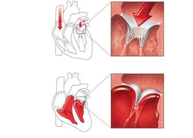
Atrioventricular (AV) Valves
AV valves prevent backflow of blood into the atria when the ventricles contract. They are anchored by chordae tendineae to papillary muscles, ensuring one-way flow.
Tricuspid Valve: Located between the right atrium and right ventricle.
Mitral (Bicuspid) Valve: Located between the left atrium and left ventricle.
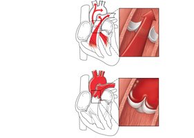
Semilunar (SL) Valves
SL valves prevent backflow into the ventricles when the ventricles relax. They open and close in response to pressure changes during the cardiac cycle.
Pulmonary Semilunar Valve: Between the right ventricle and pulmonary trunk.
Aortic Semilunar Valve: Between the left ventricle and aorta.
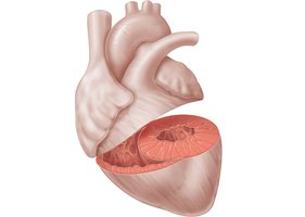
Structural Differences Between Ventricles
Ventricular Wall Thickness
The left ventricle has a much thicker wall than the right ventricle, reflecting the greater force needed to pump blood through the high-resistance systemic circuit compared to the low-resistance pulmonary circuit.
Summary Table: Heart Valves and Their Locations
Valve | Location | Function |
|---|---|---|
Tricuspid (Right AV) | Between right atrium and right ventricle | Prevents backflow into right atrium |
Mitral (Left AV/Bicuspid) | Between left atrium and left ventricle | Prevents backflow into left atrium |
Pulmonary Semilunar | Between right ventricle and pulmonary trunk | Prevents backflow into right ventricle |
Aortic Semilunar | Between left ventricle and aorta | Prevents backflow into left ventricle |
Key Concepts and Additional Information
Chordae Tendineae: Tendinous cords that anchor AV valve cusps to papillary muscles, preventing valve prolapse.
Papillary Muscles: Contract to tighten chordae tendineae during ventricular contraction.
Fibrous Skeleton of the Heart: Dense connective tissue that supports valves and electrically isolates atria from ventricles.
Example: Cardiac Cycle and Valve Operation
During ventricular contraction (systole), AV valves close to prevent backflow into the atria, while SL valves open to allow blood ejection. During ventricular relaxation (diastole), SL valves close to prevent arterial blood from re-entering the ventricles, and AV valves open to allow ventricular filling.
Equations
The cardiac output (CO) is a key measure of heart function:
Where:
CO: Cardiac Output (mL/min)
HR: Heart Rate (beats/min)
SV: Stroke Volume (mL/beat)
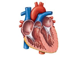
Visual Summary

 *Additional info: The above notes integrate anatomical and physiological context to provide a comprehensive overview of the heart's structure and function, suitable for ANP college students preparing for exams.*
