Back
BackThe Heart: Structure, Function, and Cardiac Physiology
Study Guide - Smart Notes
 Tailored notes based on your materials, expanded with key definitions, examples, and context.
The Heart: Structure and Circulation
Heart Anatomy Overview
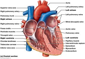
The heart is a muscular organ responsible for pumping blood throughout the body via the circulatory system. It is divided into right and left sides, each with distinct roles in pulmonary and systemic circulation.
Right heart: Receives deoxygenated blood from the body and pumps it to the lungs.
Left heart: Receives oxygenated blood from the lungs and pumps it to the rest of the body.
Circulation of Blood Through the Heart and Major Vessels
Blood flows through the heart in a specific sequence, ensuring efficient oxygenation and nutrient delivery.
Deoxygenated blood enters the right atrium via the superior and inferior vena cava.
It passes through the tricuspid valve into the right ventricle, then is pumped through the pulmonary valve into the pulmonary arteries and to the lungs.
Oxygenated blood returns via the pulmonary veins to the left atrium, passes through the mitral (bicuspid) valve into the left ventricle, and is pumped through the aortic valve into the aorta for systemic distribution.
Coronary Circulation
The coronary circulation supplies the myocardium (heart muscle) with oxygenated blood.
Arterial supply: Right and left coronary arteries originate from the aorta at the sinuses of Valsalva.
Venous drainage: Deoxygenated blood is collected by the coronary sinus and emptied into the right atrium.
Key Terms
Anastomosis: Union or joining of two blood vessels, nerves, or lymphatics, providing alternative routes for circulation.
Collateral circulation: Alternative route of blood flow to an area, often via anastomoses.
Ischemia: Insufficient blood supply to an organ or tissue, usually due to a blocked artery.
Infarction: Localized tissue necrosis resulting from obstruction of blood supply.
Angina pectoris: Chest pain resulting from myocardial ischemia.
Cardiac Muscle and Electrophysiology
Cardiomyocytes: Structure and Function
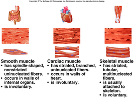
Cardiomyocytes are specialized muscle cells responsible for the heart's contractile activity.
Branched cells connected by intercalated discs (containing gap junctions and desmosomes).
Stimulus for contraction originates from pacemaker cells, regulated by the autonomic nervous system.
Action potentials last longer than in skeletal muscle, preventing tetanus.
Cells are optimized for aerobic metabolism (abundant mitochondria, myoglobin, and capillary density).
Autorhythmic and Contractile Cells
The heart contains two main types of myocytes:
Autorhythmic Cells | Contractile Cells |
|---|---|
Located in the intrinsic conduction system (SA node, AV node, AV bundle, bundle branches, Purkinje fibers) | All other myocytes |
Initiate action potentials and set heart rate | Contract and relax during systole and diastole; serve as pumping cells |
Action Potentials in Cardiac Cells
Cardiac action potentials differ between autorhythmic (pacemaker) and contractile cells.
Pacemaker potential: Gradual depolarization due to decreased K+ efflux and increased Na+ influx through slow Na+ channels.
Threshold (-40 mV): Ca2+ channels open, causing rapid depolarization.
Repolarization: Ca2+ channels close, K+ channels open, K+ leaves the cell.
Contractile cell action potential:
Depolarization: Fast Na+ channels open, Na+ enters.
Plateau: Ca2+ enters from SR and ECF, K+ permeability decreases, prolonging depolarization.
Repolarization: Ca2+ channels close, K+ channels open, K+ leaves.
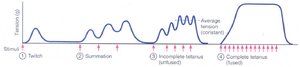
Summation and Tetanus in Muscle
Unlike skeletal muscle, cardiac muscle cannot undergo tetanus due to a long refractory period.
Skeletal muscle: Action potentials can summate, leading to tetanus.
Cardiac muscle: Refractory period nearly as long as contraction, preventing summation and allowing relaxation for chamber filling.
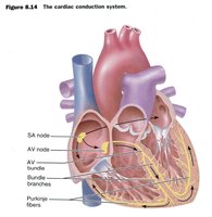
Cardiac Conduction System and ECG
Cardiac Conduction System
The heart's intrinsic conduction system coordinates contraction and ensures efficient pumping.
Key structures: SA node, AV node, AV bundle (bundle of His), bundle branches, Purkinje fibers.
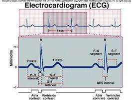
Electrocardiogram (ECG/EKG)
An ECG records the electrical activity of the heart, allowing analysis of heart rate, rhythm, and conduction abnormalities.
P wave: Atrial depolarization
QRS complex: Ventricular depolarization
T wave: Ventricular repolarization
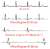
Common Arrhythmias
Arrhythmias are abnormal heart rhythms detected by ECG analysis.
Sinus tachycardia: Accelerated SA node impulses (>100 bpm).
Atrial flutter: Single ectopic pacemaker, atrial rate 200–300 bpm, saw-toothed waves, not every P wave initiates a QRS complex.
Atrial fibrillation: Multiple ectopic foci, atrial rate 450–600 bpm, irregular P waves, erratic ventricular response.
Ventricular fibrillation: Multiple ectopic foci, no effective ventricular contraction, no pulse, distorted QRS complexes.
Atrioventricular (AV) block: Impaired conduction from atria to ventricles; classified as first, second, or third degree based on severity.
The Cardiac Cycle
Phases of the Cardiac Cycle
The cardiac cycle describes the sequence of electrical and mechanical events during one heartbeat.
Atrial systole: Atria contract, pushing blood into ventricles.
Ventricular systole: Ventricles contract, ejecting blood into arteries.
Diastole: Chambers relax and fill with blood.

Electrical and Mechanical Events
Electrical events (depolarization and repolarization) precede mechanical contraction and relaxation.
P wave: Atrial depolarization (atrial systole)
QRS complex: Ventricular depolarization (ventricular systole)
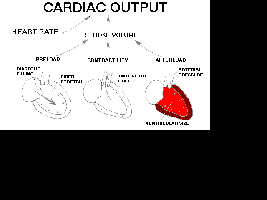
Cardiac Output and Regulation
Cardiac Output (CO)
Cardiac output is the volume of blood ejected by each ventricle per minute.
At rest, average CO is 5 L/min.
Calculated as:
Example: HR = 70 bpm, SV = 70 mL/beat → CO = 4,900 mL/min or 4.9 L/min
Stroke Volume (SV)
Stroke volume is the amount of blood ejected from the left ventricle with each beat.
Calculated as:
Typical values: EDV = 120 mL, ESV = 50 mL, SV = 70 mL
Ejection Fraction (EF)
Ejection fraction is the percentage of EDV ejected with each contraction, an important measure of cardiac function.
Normal EF > 50%; values < 50% may indicate cardiac failure.
Highly conditioned athletes may have EF > 90% during exercise.
Factors Affecting Stroke Volume
Preload: Degree of stretch of cardiac muscle fibers before contraction (Frank-Starling Law: increased preload increases force of contraction).
Contractility: Strength of contraction independent of preload (influenced by sympathetic stimulation, hormones, and drugs).
Afterload: Pressure the ventricles must overcome to eject blood (higher afterload reduces SV).
Regulation of Heart Rate
Heart rate is regulated by the autonomic nervous system, hormones, and other factors.
Positive chronotropic effects: Increase heart rate (e.g., sympathetic stimulation, epinephrine, thyroid hormone, increased temperature).
Negative chronotropic effects: Decrease heart rate (e.g., parasympathetic stimulation, acetylcholine, decreased temperature).
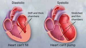
Congestive Heart Failure (CHF)
Overview
CHF occurs when the heart cannot pump blood efficiently, leading to inadequate circulation and fluid accumulation.
Causes include hypertension, arrhythmias, valve pathology, cardiomyopathy, myocardial infarction, coronary artery disease, and substance abuse.
Symptoms may include shortness of breath, edema, and fatigue.