 Back
BackThe Heart: Structure, Function, and Cardiac Cycle
Study Guide - Smart Notes
 Tailored notes based on your materials, expanded with key definitions, examples, and context.
The Pericardium and Heart Wall
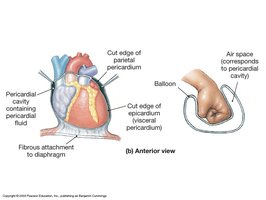
Pericardial Cavity and Pericardium
The pericardial cavity is a potential space within the thoracic cavity that surrounds the heart. It is lined by the pericardium, a double-walled sac that protects and anchors the heart, and contains pericardial fluid to reduce friction during heart movements.
Visceral pericardium (epicardium): Covers the outer surface of the heart.
Parietal pericardium: Lines the inner surface of the pericardial sac.
Fibrous pericardium: Prevents overstretching, protects, and anchors the heart to the diaphragm.
Serous pericardium: Double layer (parietal and visceral) with pericardial fluid between layers for frictionless movement.
Layers of the Heart Wall
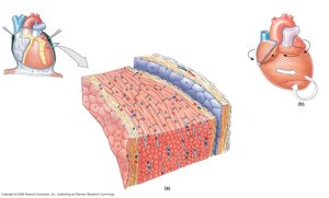
The heart wall consists of three layers, each with distinct structure and function:
Endocardium: Inner layer of simple squamous epithelium; lines the heart chambers and touches the blood.
Myocardium: Middle, thickest layer composed of cardiac muscle; responsible for contraction.
Epicardium: Outer layer, also known as the visceral layer of the serous pericardium.
Cardiac Muscle Structure and Function
Cardiac Muscle Features
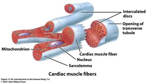
The myocardium is made of cardiac muscle, which is involuntary, striated, and branched. Cardiac muscle cells are joined end-to-end by intercalated disks, allowing the heart to contract as a functional syncytium.
Branched fibers align end-to-end.
Each cell has a single, centrally located nucleus.
Striations are present due to organized contractile proteins.
Specialized Junctions: Desmosomes and Gap Junctions
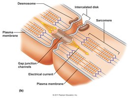
Cardiac muscle cells are connected by desmosomes (which physically join cells) and gap junctions (which allow the flow of ions and spread of action potentials). This arrangement enables rapid and coordinated contraction of the heart muscle.
Desmosomes: Provide mechanical strength by anchoring cells together.
Gap junctions: Allow electrical impulses to pass quickly between cells.
Gross Anatomy of the Heart
External and Internal Structures
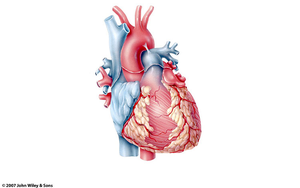
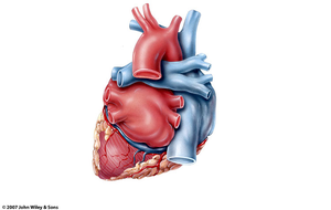
The heart consists of four chambers: right and left atria (collecting chambers) and right and left ventricles (pumping chambers). The right and left sides are separated by the muscular septum (interatrial and interventricular septa).
Atria: Receive venous blood.
Ventricles: Eject arterial blood.
Great vessels: Include the aorta, pulmonary trunk/arteries, superior and inferior vena cava, and pulmonary veins.
Heart Valves
The heart contains four main valves that ensure unidirectional blood flow:
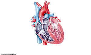
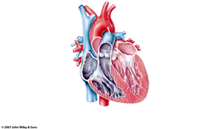
Atrioventricular (AV) valves: Tricuspid (right) and bicuspid/mitral (left) valves between atria and ventricles.
Semilunar valves: Pulmonary (right ventricle to pulmonary trunk) and aortic (left ventricle to aorta) valves.
Valve Function and Mechanism
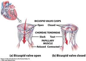
Heart valves open and close in response to pressure changes between chambers. AV valve cusps are anchored to papillary muscles by chordae tendineae, preventing inversion during contraction.
When the heart is relaxed, AV valves are open and blood flows into the ventricles.
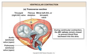
During contraction, AV valves close to prevent backflow into the atria; papillary muscles contract, tightening chordae tendineae.
Semilunar Valves
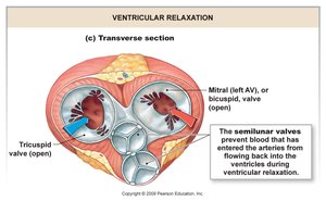
Semilunar valves prevent backflow from arteries into the ventricles during ventricular relaxation. They open when ventricular pressure exceeds arterial pressure and close when the pressure falls.
Blood Flow Through the Heart
Pathway of Blood
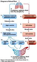
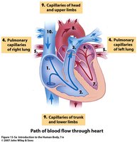
Blood flows through the heart in a specific sequence, ensuring oxygenation and systemic delivery:
Right atrium → right ventricle → pulmonary artery → lungs
Lungs → pulmonary vein → left atrium → left ventricle → aortic valve
Aortic valve → aortic arch & coronary arteries → systemic circulation
Systemic capillaries → veins → superior/inferior vena cava → right atrium
The Cardiac Cycle
Phases of the Cardiac Cycle
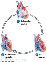
The cardiac cycle consists of alternating periods of contraction (systole) and relaxation (diastole) in the atria and ventricles. This cycle ensures efficient blood flow through the heart and to the body.
Relaxation period: All chambers are relaxed; ventricles fill passively.
Atrial systole: Atria contract, topping off ventricular filling.
Ventricular systole: Ventricles contract, ejecting blood into arteries.
Ventricular Wall Thickness
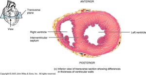
The myocardium of the left ventricle is thicker than that of the right ventricle because it must generate higher pressure to pump blood throughout the systemic circulation.
Coronary Circulation
Supplying the Heart Muscle
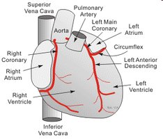
Coronary circulation supplies oxygenated blood to the myocardium. The right and left coronary arteries originate at the base of the ascending aorta and branch to nourish heart tissue. Venous blood from the myocardium returns via the coronary sinus to the right atrium.
Cardiac Conduction System
Pacemaker Cells and Electrical Pathway
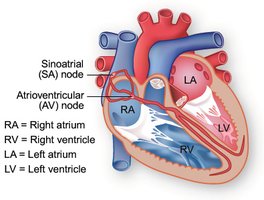
The heart's rhythmic contractions are coordinated by specialized pacemaker cells that generate and propagate action potentials. The conduction system includes:
Sinoatrial (SA) node: Natural pacemaker, initiates action potentials.
Atrioventricular (AV) node: Delays impulse, allowing atrial contraction to complete.
Bundle of His (AV bundle): Conducts impulses to ventricles.
Right and left bundle branches: Carry impulses through the interventricular septum.
Purkinje fibers: Distribute impulses through ventricular myocardium.
Electrocardiogram (ECG/EKG)
Recording Electrical Activity
An electrocardiogram (ECG or EKG) records the electrical currents generated by the heart. It is useful for detecting arrhythmias, heart damage, enlargement, and electrolyte imbalances.
P wave: Atrial depolarization
QRS complex: Ventricular depolarization
T wave: Ventricular repolarization
Cardiac Muscle Action Potential
Phases of Cardiac Muscle Action Potential
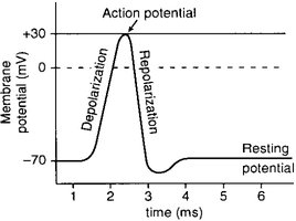
Cardiac muscle cells exhibit a unique action potential with three main phases:
Depolarization: Rapid Na+ influx through voltage-gated channels.
Plateau: Ca2+ influx balances K+ efflux, maintaining depolarization.
Repolarization: Ca2+ channels close, K+ efflux restores resting potential.
Resting membrane potential is approximately -90 mV. The plateau phase allows for sustained contraction and prevents tetanus.
Cardiac Output
Definition and Determinants
Cardiac output (CO) is the amount of blood ejected by the left ventricle per minute. It is a key indicator of the adequacy of blood flow to tissues.
Formula:
HR (Heart Rate): Number of beats per minute
SV (Stroke Volume): Volume of blood ejected per beat
Cardiac output increases with higher heart rate or stroke volume, such as during exercise or stress.
Stroke Volume Influences
Preload: Degree of ventricular stretch during filling
Afterload: Pressure the ventricles must overcome to eject blood
Contractility: Force of ventricular contraction, influenced by Ca2+ and ATP availability
Summary
This guide covers the structure and function of the heart, including the pericardium, heart wall, cardiac muscle, valves, blood flow, cardiac cycle, conduction system, and cardiac output. Mastery of these concepts is essential for understanding cardiovascular physiology and clinical applications.
