 Back
BackThe Heart: Structure, Function, and Circulation
Study Guide - Smart Notes
 Tailored notes based on your materials, expanded with key definitions, examples, and context.
The Heart: Anatomy and Location
General Features
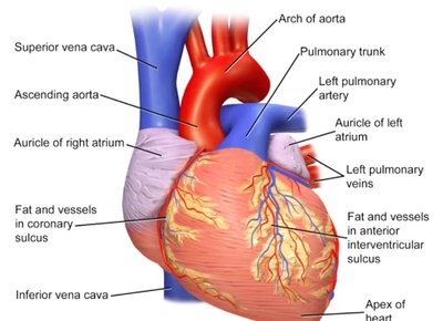
The heart is a muscular organ approximately the size of a fist, weighing between 500g and 1000g. It is centrally located in the thoracic cavity, specifically within the mediastinum and pericardial cavity, resting on the superior surface of the diaphragm and positioned between the lungs. Although it appears slightly off-center due to the left ventricle, the heart is essentially in the middle of the chest. The top of the heart is referred to as the base, while the bottom is the apex.
Mediastinum: Central compartment of the thoracic cavity.
Pericardial cavity: Fluid-filled space surrounding the heart.
Base: Superior aspect where major vessels attach.
Apex: Inferior, pointed tip of the heart.
Coverings and Layers of the Heart
The heart is protected and supported by several layers:
Pericardium: Double-walled sac with a superficial fibrous layer and a deep serous layer (parietal and visceral).
Pericardial fluid: Lubricates the heart, reducing friction during contraction.
Epicardium: Outer protective layer (visceral pericardium).
Myocardium: Muscular middle layer responsible for contraction.
Endocardium: Thin inner layer lining the chambers and valves.
Surface Features of the Heart
Grooves and Sulci
The heart's surface is marked by grooves that indicate the boundaries between chambers and house major blood vessels.
Coronary sulcus: Separates atria from ventricles.
Anterior and posterior interventricular sulci: Separate the right and left ventricles.
Coronary sinus: Collects venous blood from the heart muscle.
Chambers of the Heart
Atria: The Receiving Chambers
The heart has two atria, which receive blood returning to the heart.
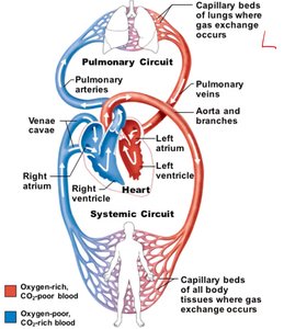
Right atrium: Receives deoxygenated blood from the body via the superior and inferior vena cava and the coronary sinus.
Left atrium: Receives oxygenated blood from the lungs via four pulmonary veins.
Auricles: Small appendages that increase atrial volume.
Pectinate muscles: Muscular ridges found in the auricles.
Ventricles: The Discharging Chambers
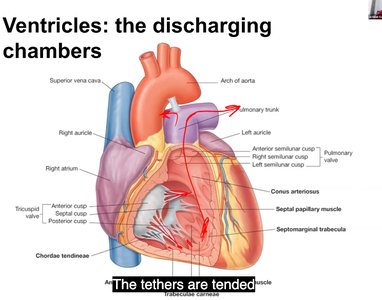
The ventricles are responsible for pumping blood out of the heart.
Right ventricle: Pumps deoxygenated blood to the lungs via the pulmonary trunk.
Left ventricle: Pumps oxygenated blood to the body via the aorta; has thicker walls due to higher force required.
Interventricular septum: Muscular wall separating the left and right ventricles.
Chordae tendineae: Tendinous cords anchoring valves to papillary muscles.
Heart Valves
Function and Types
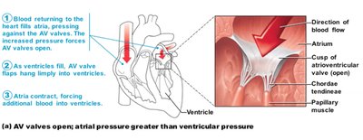
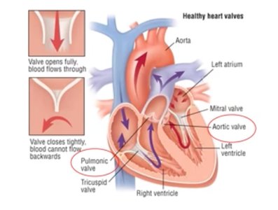
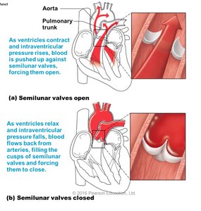
Heart valves ensure unidirectional blood flow and prevent backflow. They open and close in response to pressure changes within the chambers.
Atrioventricular (AV) valves: Located between atria and ventricles (tricuspid on the right, mitral on the left).
Semilunar (SL) valves: Located at the bases of the large arteries leaving the heart (pulmonary and aortic valves).
Valve Mechanics
AV valves open when atrial pressure exceeds ventricular pressure, allowing blood to flow into the ventricles.
AV valves close when ventricles contract, preventing backflow into the atria.
Semilunar valves open when ventricular pressure exceeds arterial pressure, allowing blood to exit the heart.
Semilunar valves close when arterial pressure exceeds ventricular pressure, preventing backflow into the ventricles.
Homeostatic Imbalance: Valve Disorders
Common Conditions
Two major conditions can severely weaken the heart:
Incompetent valve: Blood backflows, causing the heart to repump the same blood.
Valvular stenosis: Stiff valve flaps constrict the opening, requiring the heart to exert more force.
Defective valves may be replaced with mechanical, animal, or cadaver valves.
Coronary Circulation
Blood Supply to the Heart Muscle
The heart receives its own blood supply through the coronary circulation, which is the shortest in the body and accounts for about 1/20th of the total blood supply.
Blood is delivered to the heart muscle when the heart is relaxed.
The left ventricle receives most of the coronary blood supply.
Coronary arteries: Left and right coronary arteries arise from the base of the aorta and encircle the heart in the coronary sulcus.
Coronary veins: Collect deoxygenated blood from the heart muscle and empty into the right atrium via the coronary sinus.
Summary Table: Heart Chambers and Valves
Chamber | Function | Associated Valve |
|---|---|---|
Right Atrium | Receives deoxygenated blood from body | Tricuspid (AV) Valve |
Right Ventricle | Pumps blood to lungs | Pulmonary (SL) Valve |
Left Atrium | Receives oxygenated blood from lungs | Mitral (AV) Valve |
Left Ventricle | Pumps blood to body | Aortic (SL) Valve |
Key Equations
Blood Flow and Pressure
Blood flow through the heart is governed by pressure gradients:
Blood flows from areas of higher pressure to lower pressure.
Valve opening and closing is determined by relative pressures in the chambers and vessels.
Equation for blood flow:
Where Q is blood flow, ΔP is the pressure difference, and R is resistance.
Equation for cardiac output:
Where CO is cardiac output, HR is heart rate, and SV is stroke volume.
Additional info:
The heart's structure and function are essential for maintaining systemic and pulmonary circulation, ensuring oxygen delivery and waste removal throughout the body.
Valve disorders can significantly impact cardiac efficiency and may require surgical intervention.
