 Back
BackThe Heart: Structure, Function, and Circulation
Study Guide - Smart Notes
 Tailored notes based on your materials, expanded with key definitions, examples, and context.
Heart Position, Size, and Shape
Location and Orientation
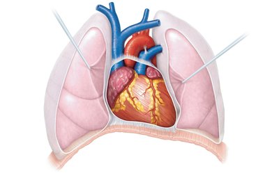
The heart is a muscular organ located in the mediastinum, the central compartment of the thoracic cavity. It is roughly the size of a closed fist. The base of the heart is positioned superiorly, while the apex points inferiorly and to the left, resting on the diaphragm. This orientation is crucial for understanding the anatomical relationships of the heart with surrounding structures such as the lungs and diaphragm.
The Pericardium and Heart Wall
Pericardium
The pericardium is a double-walled sac that encloses the heart, providing protection and reducing friction as the heart beats. It consists of two main layers:
Fibrous pericardium: The tough, outer layer that anchors the heart in place.
Serous pericardium: A thinner, inner layer that is further divided into the parietal layer (lining the internal surface of the fibrous pericardium) and the visceral layer (epicardium, which covers the heart surface).
The pericardial cavity is the space between these layers, filled with serous fluid to minimize friction.
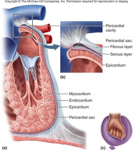
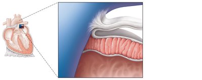
Heart Wall Structure
The heart wall is composed of three layers:
Epicardium: The outermost layer, also known as the visceral layer of the serous pericardium.
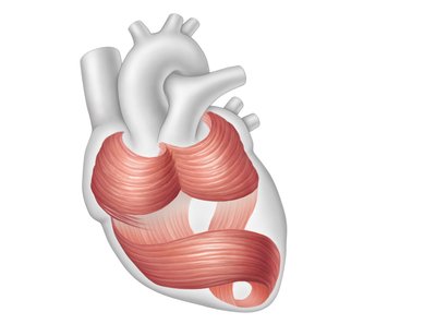
Myocardium: The thick, middle layer made of cardiac muscle tissue responsible for the heart's contractile function. The muscle fibers are arranged in a vortex pattern for efficient contraction.
Endocardium: The innermost layer, consisting of simple squamous epithelium, lining the heart chambers and covering the valves.
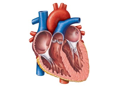
Chambers of the Heart
Atria and Ventricles
The heart contains four chambers:
Right and Left Atria: Thin-walled, superior chambers that receive blood returning to the heart. They are separated by the interatrial septum and have auricles (ear-like extensions) and pectinate muscles (internal ridges).
Right and Left Ventricles: Thick-walled, inferior chambers that pump blood out of the heart. They are separated by the interventricular septum and contain trabeculae carneae (internal ridges).
Pulmonary and Systemic Circuits
Overview of Circulation
The heart functions as a double pump, supporting two major circulatory circuits:
Pulmonary Circuit: Carries deoxygenated blood from the right heart to the lungs for gas exchange, then returns oxygenated blood to the left heart.
Systemic Circuit: Distributes oxygenated blood from the left heart to all body tissues, where oxygen is unloaded and carbon dioxide is picked up, then returns deoxygenated blood to the right heart.
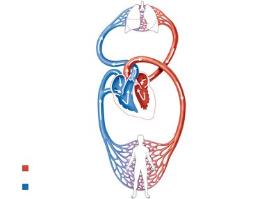
Pathway of Blood Through the Heart
Right atrium → tricuspid valve → right ventricle
Right ventricle → pulmonary semilunar valve → pulmonary trunk → pulmonary arteries → lungs
Lungs → pulmonary veins → left atrium
Left atrium → bicuspid (mitral) valve → left ventricle
Left ventricle → aortic semilunar valve → aorta → systemic circulation
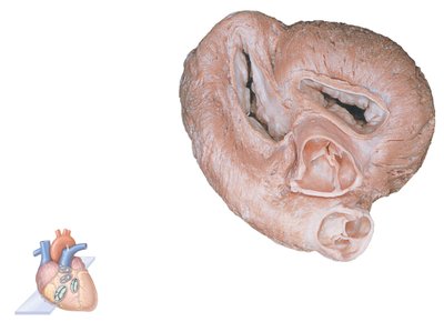
Heart Valves
Types and Functions
Heart valves ensure unidirectional blood flow through the heart:
Atrioventricular (AV) Valves: Close when ventricles contract. The right AV is the tricuspid valve; the left AV is the bicuspid (mitral) valve. Tendinous chords (chordae tendineae) tether the valves to papillary muscles.
Semilunar Valves: Close when ventricles relax. The pulmonary valve is at the exit of the right ventricle; the aortic valve is at the exit of the left ventricle.
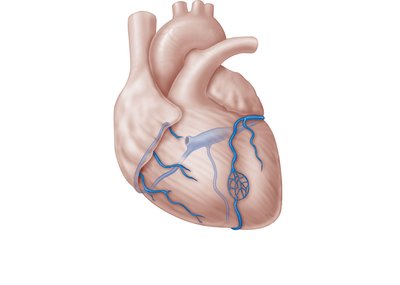
Coronary Circulation
Arterial Supply
The heart muscle (myocardium) receives its blood supply from the coronary arteries. Blockage of these arteries can lead to myocardial infarction (heart attack).
Left Coronary Artery (LCA): Branches into the anterior interventricular (left anterior descending), circumflex, and left marginal branches.
Right Coronary Artery (RCA): Branches into the right marginal and posterior interventricular branches.
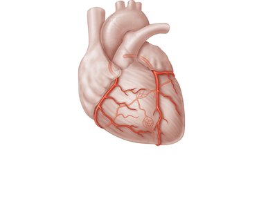
Venous Drainage
Most blood is returned to the right atrium through the coronary sinus, which receives blood from the great cardiac vein, middle cardiac vein, and left marginal vein. Small thebesian veins drain directly into the right ventricle.
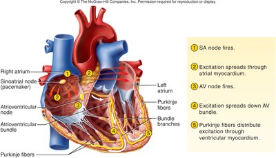
Conduction System of the Heart
Intrinsic Conduction System
The heart's rhythmic contractions are coordinated by its intrinsic conduction system, which includes:
Sinoatrial (SA) Node: The pacemaker, initiates impulses.
Atrioventricular (AV) Node: Delays the impulse, allowing atria to contract before ventricles.
AV Bundle (Bundle of His): Connects atria to ventricles.
Bundle Branches: Conduct impulses through the interventricular septum.
Purkinje Fibers: Distribute excitation through ventricular myocardium.
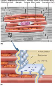
Cardiac Muscle Structure
Cardiac muscle cells (cardiocytes) are joined by intercalated discs, which contain:
Mechanical junctions: Fascia adherens and desmosomes for structural stability.
Electrical junctions: Gap junctions for rapid transmission of electrical impulses.
Nerve Supply
The autonomic nervous system modulates the heart's intrinsic activity:
Sympathetic division: Increases heart rate and force of contraction.
Parasympathetic division: Decreases heart rate, primarily via the vagus nerve.
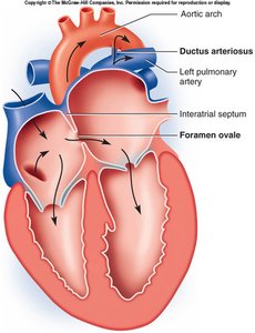
Fetal Circulation and Changes at Birth
Fetal Adaptations
In the fetal heart, the foramen ovale and ductus arteriosus allow most blood to bypass the pulmonary circuit. At birth, these structures close as the lungs inflate and resistance to blood flow decreases, forming the fossa ovalis and ligamentum arteriosum, respectively.
Heart Disease
Overview
Heart disease is the leading cause of death in the United States. Common forms include:
Coronary atherosclerosis: Can lead to myocardial infarction (heart attack).
Congenital defects: Structural abnormalities present at birth.
Myocardial hypertrophy or degeneration: Abnormal thickening or weakening of the heart muscle.
Inflammation: Of the pericardium or heart wall.
Valvular defects: Malfunction of heart valves.
Cardiac tumors: Rare growths in the heart tissue.
