 Back
BackThe Heart: Structure, Function, and Clinical Correlates
Study Guide - Smart Notes
 Tailored notes based on your materials, expanded with key definitions, examples, and context.
The Heart: Structure, Function, and Clinical Correlates
Overview of the Heart
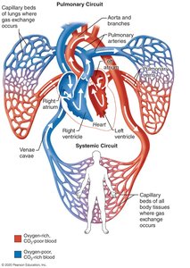
The heart is a muscular double pump essential for circulating blood throughout the body. It is divided into two main circuits: the pulmonary circuit, which sends oxygen-poor blood to the lungs, and the systemic circuit, which delivers oxygen-rich blood to the body. The heart's chambers and valves ensure unidirectional blood flow and efficient separation of oxygenated and deoxygenated blood.
Pulmonary Circuit: Right side of the heart pumps blood to the lungs for oxygenation.
Systemic Circuit: Left side of the heart pumps oxygenated blood to the rest of the body.
Atria: Receive blood from the circuits.
Ventricles: Pump blood into the circuits.
Location and Orientation of the Heart
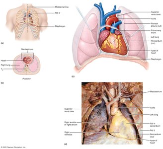
The heart is located in the mediastinum, between the lungs, with its apex pointing to the left and its base facing posteriorly. It typically weighs 250–350 grams in a healthy adult.
Four Corners of the Heart:
Superior right: 3rd rib, sternum
Inferior right: 6th rib, lateral to sternum
Superior left: 2nd rib, lateral to sternum
Inferior left: 5th intercostal space, midclavicular line
Structure of the Heart
Coverings of the Heart
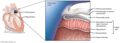
The heart is enclosed by the pericardium, which consists of two main layers:
Fibrous Pericardium: Tough, dense connective tissue that protects and anchors the heart.
Serous Pericardium: Double-layered membrane (parietal and visceral layers) that reduces friction.
Layers of the Heart Wall
Epicardium: Visceral layer of the serous pericardium.
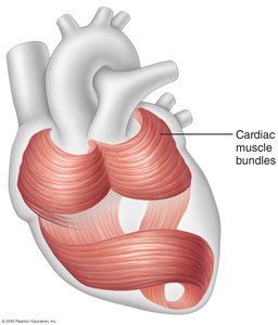
Myocardium: Cardiac muscle tissue arranged in circular and spiral bundles; responsible for contraction.
Endocardium: Endothelial lining of the heart chambers and valves.
Chambers and Internal Divisions
The heart has four chambers: two atria (superior) and two ventricles (inferior). Internal divisions include the interventricular and interatrial septa. Externally, the coronary sulcus and interventricular sulci mark chamber boundaries.
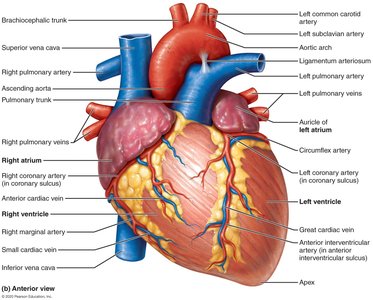
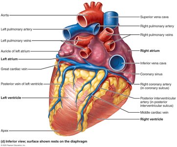
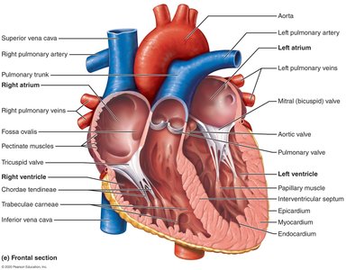
Right Atrium
Receives oxygen-poor blood from the superior and inferior venae cavae and the coronary sinus.
Contains pectinate muscles (ridges), crista terminalis (landmark), and fossa ovalis (remnant of fetal foramen ovale).
Right Ventricle
Receives blood from the right atrium via the tricuspid valve.
Pumps blood into the pulmonary trunk through the pulmonary semilunar valve.
Internal features: trabeculae carneae, papillary muscles, chordae tendineae.
Left Atrium
Receives oxygen-rich blood from the pulmonary veins.
Opens into the left ventricle via the bicuspid (mitral) valve.
Left Ventricle
Forms the apex of the heart.
Pumps blood through the aortic semilunar valve into the systemic circuit.
Internal features: trabeculae carneae, papillary muscles, chordae tendineae.
Heart Valves and Cardiac Skeleton
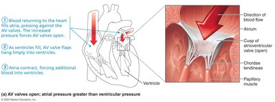
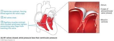
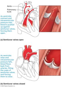
Valves ensure unidirectional blood flow. The cardiac skeleton anchors the valves and prevents overdilation, serving as an electrical insulator between atria and ventricles.
Atrioventricular (AV) Valves: Tricuspid (right), Bicuspid/Mitral (left)
Semilunar Valves: Pulmonary (right), Aortic (left)
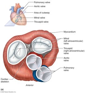
Heart Sounds
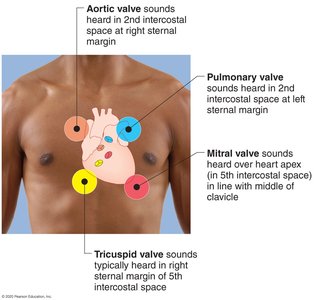
The "lub-dup" sounds correspond to valve closures:
Lub: AV valves closing
Dup: Semilunar valves closing
Each valve sound is best heard at a specific location on the thoracic surface.
Pathway of Blood Through the Heart
Blood flows through the heart in a specific sequence, ensuring separation of oxygen-poor and oxygen-rich blood. Atria contract together, followed by the ventricles.
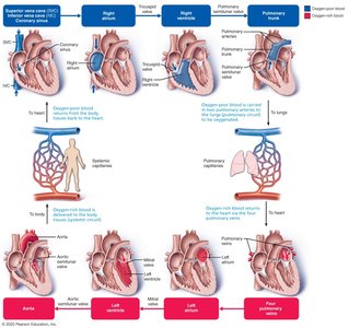
Heartbeat: Systole and Diastole
Systole: Contraction phase of a heart chamber
Diastole: Relaxation/expansion phase
Normal resting heart rate: 70–80 beats per minute
Structure of the Heart Wall
Atria have thin walls; ventricles have thick walls, especially the left ventricle, which pumps blood through the systemic circuit.
The left ventricle is three times thicker than the right and generates greater force.
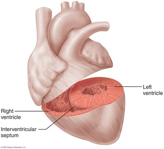
Cardiac Muscle Tissue
Microscopic Structure
Cardiac muscle tissue forms the myocardium and is striated like skeletal muscle. It contracts via the sliding filament mechanism and is composed of short, branching cells with one or two nuclei. Cells are joined by intercalated discs, which contain:
Fasciae adherens: Desmosome-like junctions for mechanical strength
Gap junctions: Allow electrical coupling between cells
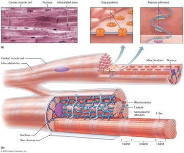
Mechanism of Contraction
Contraction is triggered by Ca2+ influx, which stimulates the sarcoplasmic reticulum to release more Ca2+.
Cardiac muscle exhibits inherent rhythmicity and can contract without neural input.
Conducting System of the Heart
Intrinsic Conduction System
The heart's conducting system consists of specialized cardiac muscle cells that generate and conduct impulses, ensuring coordinated contraction. The sinoatrial (SA) node acts as the pacemaker.
SA node → Internodal pathway → AV node → AV bundle → Bundle branches → Purkinje fibers
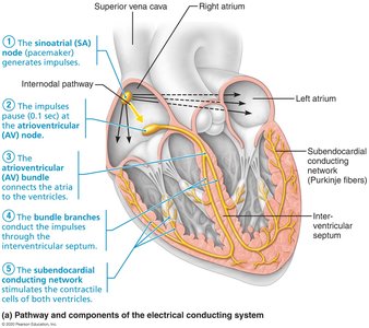
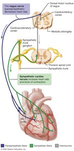
Innervation of the Heart
Heart rate and force are modulated by autonomic input:
Parasympathetic fibers (vagus nerve): Decrease heart rate; innervate SA node, AV node, and coronary arteries.
Sympathetic fibers: Increase heart rate and contraction strength; innervate SA node, AV node, coronary arteries, and cardiac muscle.
Cardiac centers in the medulla oblongata regulate autonomic input.
Blood Supply to the Heart
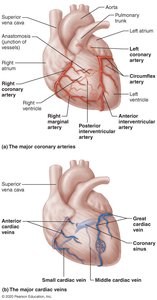
Coronary Arteries
Right and left coronary arteries arise from the base of the aorta and supply the heart wall.
Left coronary artery branches: anterior interventricular (LAD) and circumflex arteries.
Right coronary artery branches: marginal artery and posterior interventricular artery (PDA).
Cardiac Veins
Cardiac veins collect deoxygenated blood from the heart wall and drain into the coronary sinus, which empties into the right atrium.
Major veins: great cardiac vein, middle cardiac vein, small cardiac vein.
Clinical Correlates: Disorders of the Heart
Coronary Artery Disease
Atherosclerosis: Fatty deposits in coronary arteries.
Angina pectoris: Chest pain due to reduced blood flow.
Myocardial infarction: Heart attack caused by blocked coronary artery.
Silent ischemia: Reduced blood flow without pain or warning.
Heart Failure
Progressive weakening of the heart, leading to inadequate blood supply to tissues.
Congestive heart failure (CHF): Heart enlarges, pumping efficiency declines.
Pulmonary arterial hypertension: Right ventricle enlargement and potential failure.
Disorders of the Conduction System
Arrhythmias: Abnormal heart rhythms.
Ventricular fibrillation: Rapid, uncoordinated impulses in ventricles; can cause cardiac arrest.
Atrial fibrillation: Disorganized impulses in atria; increases risk of stroke.
Development and Aging of the Heart
Embryonic Development
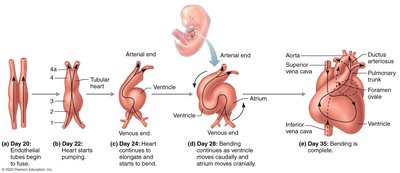
The heart begins to form and function early in embryonic development. By day 22, the heart starts pumping. The chambers develop from a simple tube, differentiating into sinus venosus, atrium, ventricle, and bulbus cordis.
Congenital Heart Defects
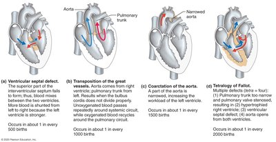
Most defects arise during the second month of development. They are classified into two main categories: defects causing inadequate oxygenation of blood and defects increasing ventricular workload.
Defect | Description | Incidence |
|---|---|---|
Ventricular septal defect | Opening in interventricular septum allows blood mixing | 1 in 500 births |
Transposition of great vessels | Aorta and pulmonary trunk are switched | 1 in 1000 births |
Coarctation of the aorta | Narrowed aorta increases left ventricular workload | 1 in 1500 births |
Tetralogy of Fallot | Four defects: pulmonary stenosis, ventricular septal defect, overriding aorta, right ventricular hypertrophy | 1 in 2000 births |
The Heart in Old Age
Heart function is generally well maintained with age.
Regular aerobic exercise strengthens the heart and helps clear fatty deposits.
Age-related changes include valve thickening, reduced cardiac reserve, and fibrosis of cardiac muscle.
