 Back
BackThe Heart: Structure, Function, and Clinical Relevance
Study Guide - Smart Notes
 Tailored notes based on your materials, expanded with key definitions, examples, and context.
The Heart: Structure and Function
Overview of the Heart
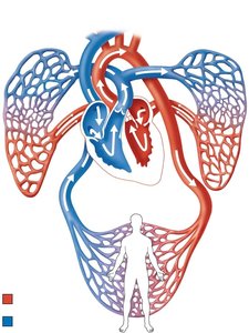
The heart is a muscular organ responsible for pumping blood throughout the body via two main circuits: the pulmonary and systemic circuits. It is located in the mediastinum of the thoracic cavity, between the lungs and above the diaphragm.
Pulmonary Circuit: Carries blood between the heart and lungs for gas exchange.
Systemic Circuit: Delivers oxygenated blood from the heart to the rest of the body and returns deoxygenated blood back to the heart.
Location and Orientation of the Heart
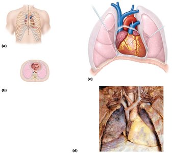
The heart lies obliquely in the thoracic cavity, with its base directed toward the right shoulder and its apex pointing to the left hip. It is protected by the rib cage and rests on the superior surface of the diaphragm.
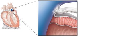
Layers of the Heart Wall and Pericardium
The heart wall consists of three layers, and the heart is enclosed in a double-walled sac called the pericardium.
Epicardium: The outermost layer, also known as the visceral layer of the serous pericardium.
Myocardium: The thick, middle layer composed of cardiac muscle responsible for contraction.
Endocardium: The innermost layer lining the heart chambers and valves.
Pericardium: Includes the fibrous pericardium (outer) and serous pericardium (parietal and visceral layers).
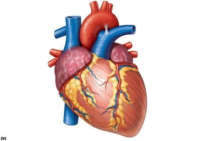
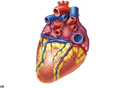
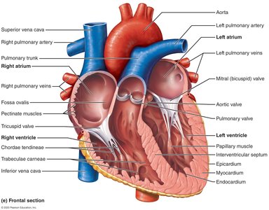
Gross Anatomy of the Heart
The heart has four chambers: two atria (upper chambers) and two ventricles (lower chambers). Major vessels enter and leave the heart at its base.
Right Atrium: Receives deoxygenated blood from the body via the superior and inferior vena cava.
Right Ventricle: Pumps blood to the lungs via the pulmonary trunk.
Left Atrium: Receives oxygenated blood from the lungs via the pulmonary veins.
Left Ventricle: Pumps oxygenated blood to the body via the aorta.
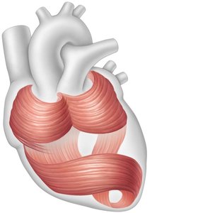
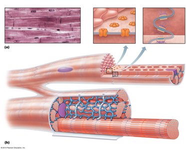
Cardiac Muscle Structure
The myocardium is composed of cardiac muscle cells arranged in circular and spiral bundles, allowing efficient contraction and ejection of blood.
Cardiac Muscle Cells: Striated, branched, and interconnected by intercalated discs containing gap junctions and fasciae adherens.
Function: Enables coordinated contraction for effective pumping action.
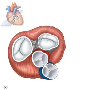
Heart Valves and Blood Flow
Heart Valves
The heart contains four main valves that ensure unidirectional blood flow:
Atrioventricular (AV) Valves: Tricuspid (right) and mitral/bicuspid (left) valves between atria and ventricles.
Semilunar Valves: Pulmonary (right) and aortic (left) valves at the exits of the ventricles.
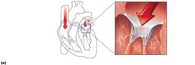
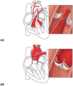
Function of the Atrioventricular Valves
AV valves prevent backflow of blood into the atria during ventricular contraction.
When atrial pressure exceeds ventricular pressure, AV valves open, allowing blood to flow into the ventricles.
When ventricles contract, AV valves close, and chordae tendineae with papillary muscles prevent valve prolapse.
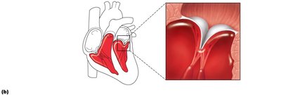
Function of the Semilunar Valves
Semilunar valves prevent backflow of blood from the arteries into the ventricles after contraction.
Open when ventricular pressure exceeds arterial pressure during systole.
Close when ventricles relax and arterial pressure exceeds ventricular pressure.
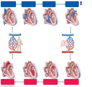
Blood Flow Through the Heart
Blood flows through the heart in a specific sequence, passing through all four chambers and valves:
Deoxygenated blood enters the right atrium from the superior and inferior vena cava.
Passes through the tricuspid valve into the right ventricle.
Pumped through the pulmonary valve into the pulmonary trunk and arteries to the lungs.
Oxygenated blood returns via pulmonary veins to the left atrium.
Passes through the mitral valve into the left ventricle.
Pumped through the aortic valve into the aorta and systemic circulation.
Electrical Conducting System of the Heart
Intrinsic Conducting System
The heart's intrinsic conducting system coordinates the heartbeat, ensuring efficient contraction of the atria and ventricles.
Sinoatrial (SA) Node: Pacemaker of the heart, initiates electrical impulses.
Atrioventricular (AV) Node: Delays impulse, allowing atrial contraction before ventricular contraction.
AV Bundle (Bundle of His): Only electrical connection between atria and ventricles.
Bundle Branches: Conduct impulses through the interventricular septum.
Purkinje Fibers: Stimulate ventricular muscle cells for contraction.
*Additional info: The delay at the AV node ensures that the atria have ejected their blood into the ventricles before the ventricles contract.*
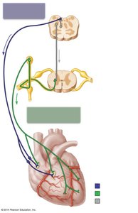
Autonomic Innervation of the Heart
The heart rate and force of contraction are modulated by the autonomic nervous system:
Sympathetic Stimulation: Increases heart rate and contractility via cardiac nerves.
Parasympathetic Stimulation: Decreases heart rate via the vagus nerve.
Clinical Scenarios and Applications
Atrial Fibrillation
Atrial fibrillation is an arrhythmia affecting the atria, leading to irregular and often rapid heart rhythm. It can cause inefficient blood flow and increase the risk of stroke and heart failure.
Affected Chambers: Both right and left atria.
Potential Complications: Thrombus formation, embolic stroke, heart failure.
Postural Orthostatic Tachycardia Syndrome (POTS)
POTS is characterized by an excessive increase in heart rate upon standing, often accompanied by dizziness, palpitations, and fatigue. Symptoms improve when lying down.
Diagnosis: Based on heart rate and blood pressure changes with position.
Treatment: Increased fluid and salt intake, physical counter-maneuvers, medications as needed.
Heart Sounds and Auscultation
Heart sounds are produced by the closing of valves. The first heart sound (S1) is due to AV valve closure, and the second heart sound (S2) is due to semilunar valve closure. Auscultation is used to assess heart function and detect abnormalities.
S1: Closure of mitral and tricuspid valves.
S2: Closure of aortic and pulmonary valves.
Summary Table: Heart Valves and Their Locations
Valve | Location | Function |
|---|---|---|
Tricuspid (Right AV) | Between right atrium and right ventricle | Prevents backflow into right atrium |
Mitral (Left AV) | Between left atrium and left ventricle | Prevents backflow into left atrium |
Pulmonary Semilunar | Between right ventricle and pulmonary trunk | Prevents backflow into right ventricle |
Aortic Semilunar | Between left ventricle and aorta | Prevents backflow into left ventricle |
Key Terms
Myocardium: Muscular layer of the heart wall.
Pericardium: Double-walled sac enclosing the heart.
SA Node: Pacemaker of the heart.
AV Node: Electrical relay station between atria and ventricles.
Chordae Tendineae: Tendinous cords anchoring AV valve cusps to papillary muscles.
