 Back
BackThe Heart: Structure, Function, and Physiology
Study Guide - Smart Notes
 Tailored notes based on your materials, expanded with key definitions, examples, and context.
The Heart: Structure, Function, and Physiology
Overview of the Cardiovascular System
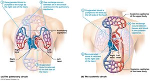
The cardiovascular system is responsible for transporting blood, nutrients, gases, and wastes throughout the body. The heart is the central organ of this system, functioning as a muscular pump that maintains blood circulation through two major circuits: the pulmonary and systemic circuits.
Pulmonary circuit: Carries deoxygenated blood from the right side of the heart to the lungs and returns oxygenated blood to the left side of the heart.
Systemic circuit: Delivers oxygenated blood from the left side of the heart to the body and returns deoxygenated blood to the right side.
Location and Basic Structure of the Heart
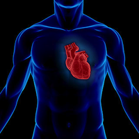
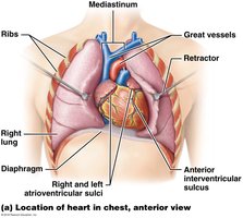
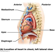
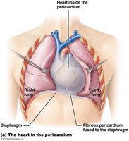
The heart is located in the mediastinum, the central compartment of the thoracic cavity, between the lungs and above the diaphragm. It is roughly the size of a fist and is oriented with its apex pointing downward and to the left.
Mediastinum: The anatomical region between the lungs where the heart resides.
Apex: The pointed end of the heart, directed inferiorly and to the left.
Base: The broad, superior portion where major vessels attach.
Functions of the Heart
The heart serves as a pump to propel blood through the pulmonary and systemic circuits. Additionally, it acts as an endocrine organ by releasing hormones such as atrial natriuretic peptide (ANP) to help regulate blood pressure.
Pericardium and Heart Wall
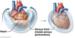
The heart is enclosed in a double-walled sac called the pericardium, which protects and anchors the heart while allowing it to move freely during contraction.
Fibrous pericardium: Tough outer layer that prevents overexpansion.
Serous pericardium: Thin, double-layered membrane (parietal and visceral layers) with serous fluid in between to reduce friction.
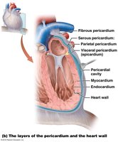
Epicardium (visceral pericardium): Outer layer, contains blood vessels and fat.
Myocardium: Thick, muscular middle layer responsible for contraction.
Endocardium: Smooth inner lining that reduces friction and is continuous with blood vessels.
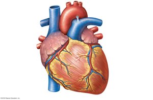
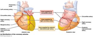
Coronary Circulation
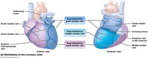
The heart muscle (myocardium) requires its own blood supply, provided by the coronary arteries and drained by the cardiac veins. This system ensures the heart receives adequate oxygen and nutrients to sustain its activity.
Left and right coronary arteries: Branch from the aorta and supply the heart muscle.
Cardiac veins and coronary sinus: Collect deoxygenated blood from the myocardium and return it to the right atrium.
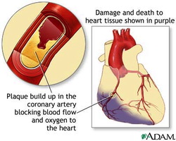
Clinical Application: Angina and Myocardial Infarction
Angina pectoris: Chest pain due to partial obstruction of coronary blood flow, leading to ischemia and lactic acid buildup.
Myocardial infarction (MI): Heart attack caused by complete blockage of a coronary artery, resulting in death of cardiac tissue.
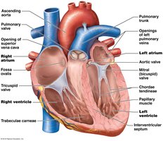
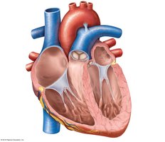
Chambers and Septa of the Heart
The heart contains four chambers: two atria (upper chambers) and two ventricles (lower chambers). Septa separate the right and left sides to prevent mixing of oxygenated and deoxygenated blood.
Right atrium: Receives deoxygenated blood from the body.
Left atrium: Receives oxygenated blood from the lungs.
Right ventricle: Pumps blood to the lungs.
Left ventricle: Pumps blood to the systemic circulation.
Interatrial septum: Separates the atria.
Interventricular septum: Separates the ventricles.
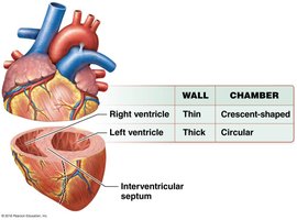
Structural Differences Between Ventricles
Left ventricle: Thicker wall, pumps blood to the entire body (high resistance).
Right ventricle: Thinner wall, pumps blood to the lungs (low resistance).
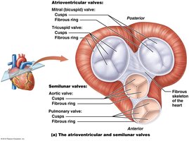
Heart Valves
Heart valves ensure unidirectional blood flow through the heart. There are two types: atrioventricular (AV) valves and semilunar valves.
Right AV (tricuspid) valve: Between right atrium and ventricle.
Left AV (mitral/bicuspid) valve: Between left atrium and ventricle.
Chordae tendineae: Tendinous cords that anchor AV valves to papillary muscles, preventing prolapse.
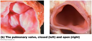
Pulmonary semilunar valve: Between right ventricle and pulmonary trunk.
Aortic semilunar valve: Between left ventricle and aorta.
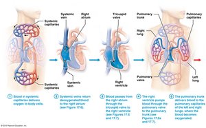
Blood Flow Through the Heart
Blood flows through the heart in a specific sequence, passing through each chamber and valve to ensure efficient circulation.
Deoxygenated blood enters the right atrium via the superior and inferior vena cava.
Passes through the tricuspid valve into the right ventricle.
Pumped through the pulmonary semilunar valve into the pulmonary trunk and arteries to the lungs.
Oxygenated blood returns via pulmonary veins to the left atrium.
Passes through the mitral valve into the left ventricle.
Pumped through the aortic semilunar valve into the aorta and systemic circulation.
Cardiac Muscle Tissue Anatomy and Electrophysiology
Types of Cardiac Cells
Pacemaker cells: Specialized cells (~1%) that spontaneously generate action potentials, setting the heart's rhythm.
Contractile cells: The majority (~99%) of cardiac cells, responsible for the forceful contraction of the heart.
Autorhythmicity: The heart's ability to generate its own rhythm without external nervous input.
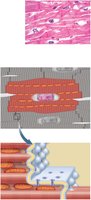
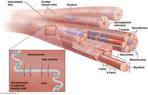
Histology of Cardiac Muscle
Cardiac muscle cells (cardiocytes) are striated, branched, and interconnected by intercalated discs, which are unique to cardiac tissue.
Intercalated discs: Specialized junctions containing interdigitating folds, mechanical junctions (fascia adherens and desmosomes), and electrical junctions (gap junctions).
Gap junctions: Allow ions to flow between cells, enabling coordinated contraction.
Desmosomes: Provide mechanical strength, preventing cells from pulling apart during contraction.


Electrophysiology of Cardiac Muscle
Voltage: Difference in electrical potential between two points.
Membrane potential: Voltage across the cell membrane.
Resting membrane potential: Typically between -60 and -90 mV in cardiac cells.
Depolarization: Membrane potential becomes less negative as positive ions enter the cell.
Repolarization: Return to resting membrane potential as positive ions leave the cell.
Action Potentials in Cardiac Cells
Contractile cell action potential: Characterized by a rapid depolarization, a plateau phase (due to Ca2+ influx), and repolarization.
Plateau phase: Prolongs the action potential, allowing for sustained contraction and efficient blood ejection.
Pacemaker cell action potential: Lacks a stable resting potential; spontaneous depolarization leads to rhythmic firing.
Key equation (Nernst equation for equilibrium potential):
Additional info: The plateau phase is unique to cardiac muscle and is essential for preventing tetanus, ensuring rhythmic contractions.
The Cardiac Conduction System
The conduction system coordinates the heartbeat, ensuring efficient and synchronized contraction of the atria and ventricles.
Sinoatrial (SA) node: Pacemaker of the heart, initiates each heartbeat.
Atrioventricular (AV) node: Electrical gateway to the ventricles, delays the impulse to allow ventricular filling.
AV bundle (bundle of His): Conducts impulses from the AV node to the ventricles.
Purkinje fibers: Distribute the impulse throughout the ventricular myocardium.
Nerve Supply to the Heart
Sympathetic nerves: Increase heart rate and contraction strength.
Parasympathetic nerves (vagus nerve): Decrease heart rate.
The Electrocardiogram (ECG/EKG)
An ECG records the electrical activity of the heart and is used to diagnose arrhythmias and other cardiac conditions.
P wave: Atrial depolarization.
QRS complex: Ventricular depolarization.
T wave: Ventricular repolarization.
Cardiac Rhythm and Arrhythmias
Sinus rhythm: Normal rhythm set by the SA node (60–100 bpm).
Ectopic focus: Abnormal pacemaker site outside the SA node.
Arrhythmia: Any abnormal cardiac rhythm (e.g., heart block, premature ventricular contractions, ventricular fibrillation).
Mechanical Physiology of the Heart: The Cardiac Cycle
Phases of the Cardiac Cycle
The cardiac cycle consists of a series of events that occur during one heartbeat, including contraction (systole) and relaxation (diastole) of the atria and ventricles.
Systole: Contraction phase, blood is ejected from the chambers.
Diastole: Relaxation phase, chambers fill with blood.
Valve Function During the Cardiac Cycle
AV valves open during ventricular diastole and close during systole.
Semilunar valves open during ventricular systole and close during diastole.
Heart Sounds
S1 ("lub"): Closure of AV valves at the beginning of ventricular systole.
S2 ("dup"): Closure of semilunar valves at the beginning of ventricular diastole.
Pressure and Volume Changes
Pressure changes in the heart chambers drive the opening and closing of valves and the movement of blood. The left ventricle generates higher pressures than the right due to the greater resistance of the systemic circuit.
Connecting Electrical and Mechanical Events
The electrical events (as seen on the ECG) precede and trigger the mechanical events of the cardiac cycle, ensuring coordinated contraction and efficient blood flow.
Phase | Event | Valve Status |
|---|---|---|
Ventricular filling | Ventricles fill with blood | AV open, SL closed |
Isovolumetric contraction | Ventricles contract, pressure rises | AV closed, SL closed |
Ventricular ejection | Blood ejected into arteries | AV closed, SL open |
Isovolumetric relaxation | Ventricles relax, pressure falls | AV closed, SL closed |
Additional info: The cardiac cycle is tightly regulated to maintain adequate tissue perfusion and respond to changes in metabolic demand.
