 Back
BackThe Heart: Structure, Function, and Physiology
Study Guide - Smart Notes
 Tailored notes based on your materials, expanded with key definitions, examples, and context.
The Heart: Structure, Function, and Physiology
Functions of the Heart
The heart is a muscular organ responsible for pumping blood throughout the body, maintaining blood pressure, and ensuring the delivery of oxygen and nutrients to tissues while removing waste products.
Pumping Blood: The heart circulates blood through the pulmonary and systemic circuits.
Blood Pressure Regulation: The heart generates the force necessary to move blood through the vessels.
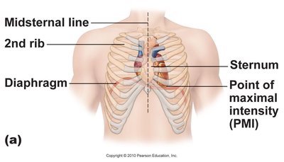
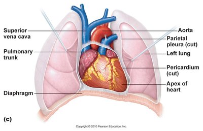
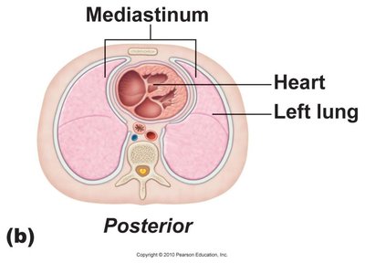
Location and Orientation of the Heart
The heart is located in the mediastinum, the central compartment of the thoracic cavity, between the lungs. Its apex rests on the diaphragm, and its base is the site where major vessels exit the heart.
Apex: Points downward and to the left, resting on the diaphragm.
Base: Superior aspect where large vessels attach.
Borders: Anterior (right ventricle), left (left ventricle), right (right atrium).
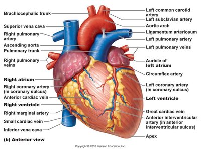
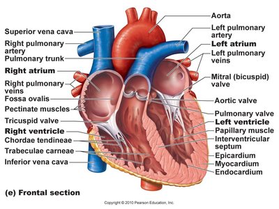
Gross Anatomy of the Heart
The heart consists of four chambers: two atria (receiving chambers) and two ventricles (discharging chambers). The right side pumps blood to the lungs (pulmonary circuit), while the left side pumps blood to the rest of the body (systemic circuit).
Right Atrium: Receives deoxygenated blood from the body.
Right Ventricle: Pumps blood to the lungs.
Left Atrium: Receives oxygenated blood from the lungs.
Left Ventricle: Pumps blood to the systemic circulation.
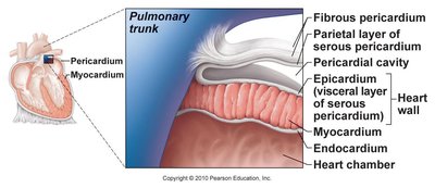
Layers of the Heart Wall
The heart wall is composed of three layers, each with distinct structure and function:
Pericardium: A double-walled sac that surrounds and protects the heart. It consists of:
Fibrous Pericardium: Dense connective tissue that anchors and protects the heart.
Serous Pericardium: Includes the parietal layer (lining the fibrous pericardium) and the visceral layer (epicardium, covering the heart surface). The pericardial cavity between these layers contains lubricating fluid.
Myocardium: The thick, muscular middle layer responsible for contraction. Composed of striated cardiac muscle cells with intercalated discs, abundant mitochondria, and rich in myoglobin and glycogen.
Endocardium: The inner lining of the heart, made of simple squamous epithelium, also covering the heart valves.
Heart Valves and Chambers
The heart contains four main valves that ensure unidirectional blood flow:
Atrioventricular (AV) Valves: Tricuspid (right) and mitral/bicuspid (left) valves separate atria from ventricles.
Semilunar Valves: Pulmonary (right ventricle to pulmonary artery) and aortic (left ventricle to aorta) valves prevent backflow into the ventricles.

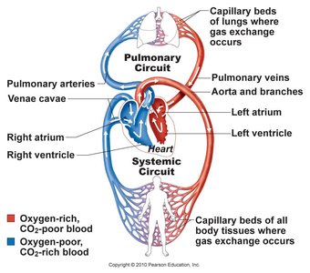
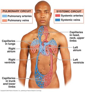
Circulatory Pathways: Pulmonary and Systemic Circuits
The heart acts as a double pump, maintaining two separate circuits:
Pulmonary Circuit: Right heart pumps deoxygenated blood to the lungs for gas exchange.
Systemic Circuit: Left heart pumps oxygenated blood to the body tissues.
Pathway of Blood Through the Heart
Blood flows through the heart in a specific sequence, passing through valves and chambers:
Superior & Inferior Vena Cava and coronary sinus → Right atrium
Right atrium → Tricuspid valve → Right ventricle
Right ventricle → Pulmonary semilunar valve → Pulmonary trunk → Pulmonary arteries → Lungs
Lungs → Pulmonary veins → Left atrium
Left atrium → Bicuspid (mitral) valve → Left ventricle
Left ventricle → Aortic semilunar valve → Aorta → Systemic circulation
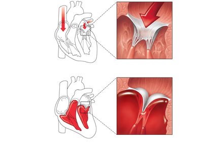
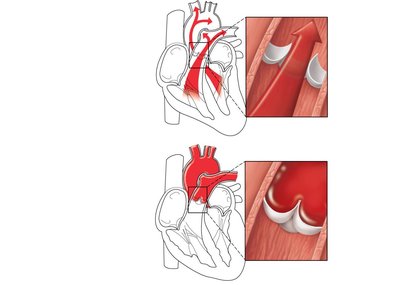
Heart Valves: Function and Mechanism
Heart valves open and close in response to pressure changes, ensuring one-way blood flow:
AV Valves: Open when atrial pressure exceeds ventricular pressure; close when ventricles contract.
Semilunar Valves: Open when ventricular pressure exceeds arterial pressure; close when ventricles relax.
Coronary Circulation
The heart muscle (myocardium) receives its own blood supply via the coronary arteries:
Right Coronary Artery (RCA): Supplies right atrium and ventricle.
Left Coronary Artery (LCA): Supplies left atrium, left ventricle, and part of right ventricle.
Venous Drainage: Cardiac veins drain into the coronary sinus, which empties into the right atrium.
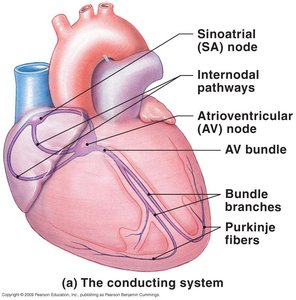
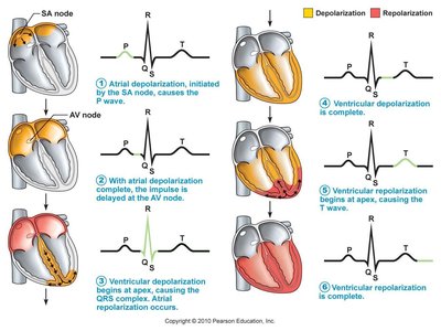
Conduction System of the Heart
The heart's electrical system coordinates contraction:
SA Node (Sinoatrial): Pacemaker, initiates action potentials (~72/min).
AV Node (Atrioventricular): Delays impulse, allowing atrial contraction before ventricular contraction.
Bundle of His, Bundle Branches, Purkinje Fibers: Rapidly conduct impulses through ventricles.
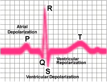
Electrocardiogram (EKG/ECG)
An EKG records the electrical activity of the heart, showing characteristic waves:
P Wave: Atrial depolarization
QRS Complex: Ventricular depolarization (and atrial repolarization)
T Wave: Ventricular repolarization
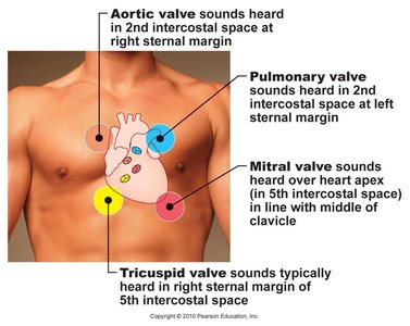
Heart Sounds and Auscultation
Heart sounds are produced by valve closure:
First sound ("Lubb"): Closure of AV valves at the start of ventricular systole.
Second sound ("Dubb"): Closure of semilunar valves at the start of ventricular diastole.
Murmurs: Abnormal sounds due to turbulent blood flow, often from valve defects.
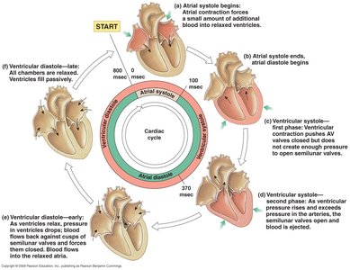
The Cardiac Cycle
The cardiac cycle describes the sequence of events in one heartbeat, including systole (contraction) and diastole (relaxation):
Ventricular Filling (Diastole): AV valves open, ventricles fill passively (~70%), then actively by atrial contraction (~30%).
Ventricular Systole: Isovolumetric contraction (all valves closed), followed by ejection phase (semilunar valves open).
Ventricular Diastole: Isovolumetric relaxation (all valves closed), then passive and active filling resumes.
Cardiac Output and Regulation
Cardiac output (CO) is the volume of blood pumped by one ventricle per minute:
Formula:
Stroke Volume (SV): Volume of blood ejected per beat (SV = EDV - ESV).
End-Diastolic Volume (EDV): Blood in ventricle after diastole (~120 mL).
End-Systolic Volume (ESV): Blood left after systole (~50 mL).
Cardiac Reserve: Difference between resting and maximal CO.
Regulation of Cardiac Output
Preload: Degree of stretch of cardiac muscle (related to venous return).
Afterload: Pressure ventricles must overcome to eject blood (arterial pressure).
Autonomic Regulation: Sympathetic increases HR and contractility; parasympathetic decreases HR.
Hormonal Regulation: Epinephrine increases HR and contractility.
Clinical Correlations
Pericarditis: Inflammation of the pericardium, can restrict heart movement.
Endocarditis: Inflammation of the endocardium, often affecting valves.
Myocardial Infarction: Death of heart muscle due to prolonged ischemia (often from atherosclerosis).
Congestive Heart Failure: Inability of the heart to pump effectively, leading to fluid accumulation in tissues (right-sided) or lungs (left-sided).
