 Back
BackThe Heart: Structure, Function, and Physiology
Study Guide - Smart Notes
 Tailored notes based on your materials, expanded with key definitions, examples, and context.
The Heart: Structure and Function
Location and Role of the Heart
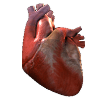
The heart is a muscular organ responsible for generating the force necessary to propel blood through the circulatory system. It is located within the mediastinum of the thorax, bordered laterally by the lungs, posteriorly by the vertebral column, and anteriorly by the sternum.
Propulsive Force: The heart acts as a pump, ensuring unidirectional blood flow via one-way valves.
Circulatory Pathways: The heart supports two main circuits: the pulmonary circuit (to the lungs) and the systemic circuit (to the rest of the body).
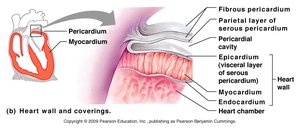
Heart Wall and Coverings
Layers of the Heart Wall
The heart wall consists of three primary layers, each with distinct structural and functional roles:
Epicardium: The outermost layer, also known as the visceral layer of the serous pericardium.
Myocardium: The thick, muscular middle layer responsible for contraction and force generation.
Endocardium: The innermost layer lining the heart chambers and valves.
The heart is enclosed by the pericardium, which consists of a fibrous outer layer and a serous inner layer, providing protection and reducing friction during heart movements.
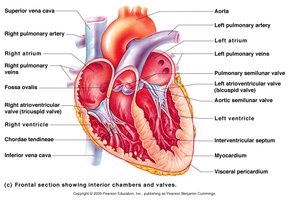
Gross Anatomy of the Heart
Chambers and Valves
The heart contains four chambers: two atria (upper chambers) and two ventricles (lower chambers). Blood flows through these chambers in a specific sequence, regulated by four main valves:
Atrioventricular (AV) Valves: Tricuspid (right) and bicuspid/mitral (left) valves prevent backflow into the atria.
Semilunar Valves: Pulmonary (right) and aortic (left) valves prevent backflow into the ventricles.
Major vessels such as the aorta, pulmonary arteries, and veins are connected to the heart, facilitating blood transport to and from the lungs and body.
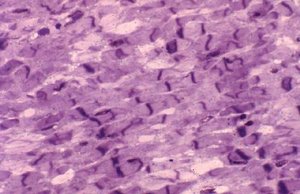
Microscopic Anatomy of Heart Muscle Tissue
Cardiac Muscle Structure
Cardiac muscle tissue is composed of specialized, striated muscle cells called cardiomyocytes. These cells are interconnected by intercalated discs, which allow rapid transmission of electrical impulses and synchronized contraction.
Striations: Indicate organized contractile proteins.
Intercalated Discs: Specialized junctions for electrical and mechanical connectivity.
Automaticity: Cardiac muscle can depolarize spontaneously without external stimuli.
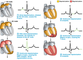
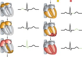
Intrinsic Conduction System and Electrocardiography
Electrical Activity of the Heart
The heart's intrinsic conduction system coordinates the rhythmic contraction of cardiac muscle. Key components include the sinoatrial (SA) node, atrioventricular (AV) node, bundle of His, bundle branches, and Purkinje fibers.
SA Node: The primary pacemaker, initiates depolarization.
AV Node: Delays the impulse, allowing atrial contraction before ventricular contraction.
Electrocardiogram (ECG/EKG): A recording of the heart's electrical activity, showing three main waves:
P wave: Depolarization of the atria (SA node activity).
QRS complex: Ventricular depolarization (and atrial repolarization).
T wave: Ventricular repolarization.
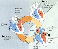
The Cardiac Cycle
Phases of the Cardiac Cycle
The cardiac cycle describes the sequence of events during one heartbeat, including contraction (systole) and relaxation (diastole) of the atria and ventricles.
Atrial Systole: Atria contract simultaneously, pushing blood into the ventricles.
Ventricular Systole: Ventricles contract, ejecting blood into the pulmonary and systemic circuits.
Diastole: Both atria and ventricles relax, allowing chambers to refill with blood.
Valves open and close in response to pressure changes, ensuring unidirectional blood flow.
Cardiovascular Physiology
Key Terms and Concepts
Understanding the physiological properties of cardiac muscle and heart function is essential for interpreting normal and abnormal heart rhythms.
Automaticity: The ability of cardiac cells to depolarize spontaneously.
Rhythmicity: The regular, continuous pattern of depolarization and repolarization.
Arrhythmia: Any irregularity or loss of rhythm in the heartbeat.
Cardiac Muscle Cannot Be Tetanized: Due to its refractory period, cardiac muscle cannot sustain a prolonged contraction.
Autonomic Nervous System (ANS): Controls heart rate via the vagus nerve; the parasympathetic division slows heart rate.
Temperature: Influences cardiac muscle function and heart rate.
Example: During exercise, sympathetic stimulation increases heart rate and contractility, while parasympathetic stimulation (via the vagus nerve) slows the heart rate during rest.
Additional info: The cardiac cycle duration is typically about 0.8 seconds in a resting adult, with systole and diastole phases ensuring efficient blood flow and oxygen delivery.
