 Back
BackThe Heart: Structure, Function, and Physiology
Study Guide - Smart Notes
 Tailored notes based on your materials, expanded with key definitions, examples, and context.
The Heart: Structure, Function, and Physiology
Function of the Heart
The heart is a muscular organ responsible for pumping blood throughout the body, supplying tissues with oxygen and nutrients while removing waste products.
Pumping blood: Ensures continuous circulation to meet metabolic demands.
Oxygen and nutrient delivery: Blood transports essential substances to cells.
Chambers of the Heart
The heart consists of four chambers: two atria and two ventricles, each with distinct roles in blood circulation.
Atria: Receive blood from veins. The right atrium receives deoxygenated blood; the left atrium receives oxygenated blood.
Ventricles: Pump blood out through arteries. The right ventricle sends blood to the lungs; the left ventricle sends blood to the body.
Veins: Return blood to the heart.
Arteries: Carry blood away from the heart.
Capillaries: Serve as exchange vessels between arteries and veins.
Pulmonary and Systemic Circuits
The heart operates two major circulatory circuits: pulmonary and systemic.
Pulmonary circuit: Carries deoxygenated blood from the right ventricle to the lungs and returns oxygenated blood to the left atrium.
Systemic circuit: Transports oxygenated blood from the left ventricle to the body and returns deoxygenated blood to the right atrium.
Pericardial Cavity and Heart Location
The heart is located in the pericardial cavity within the mediastinum, surrounded by the pericardium.
Fibrous pericardium: Lines the mediastinum.
Serous pericardium: Lines the heart and produces pericardial fluid to reduce friction.
Layers of the Heart Wall
The heart wall is composed of three layers, each with specialized functions.
Epicardium: Outer layer, also the visceral layer of the serous pericardium.
Myocardium: Middle layer, made of cardiac muscle tissue.
Endocardium: Inner layer, a simple squamous epithelium continuous with blood vessels.
Cardiac Muscle Tissue
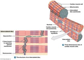
Cardiac muscle tissue is unique, enabling the heart to contract rhythmically and efficiently.
Many mitochondria: Support high energy demands.
Autorhythmicity: Can contract without nervous system stimulation.
Intercalated discs: Specialized junctions connecting cells, containing gap junctions for direct electrical communication.
Functional syncytium: Cardiac muscle functions as a single unit.
The Great Vessels
The heart is connected to major blood vessels that facilitate circulation.
Superior and inferior vena cava: Return deoxygenated blood to the right atrium.
Pulmonary trunk and arteries: Carry blood from the right ventricle to the lungs.
Pulmonary veins: Return oxygenated blood from the lungs to the left atrium.
Aorta: Delivers oxygenated blood from the left ventricle to the body.
Valves of the Heart
Valves ensure unidirectional blood flow and prevent backflow.
Atrioventricular (AV) valves:
Right AV (tricuspid) valve: Between right atrium and right ventricle.
Left AV (mitral/bicuspid) valve: Between left atrium and left ventricle.
Semilunar valves:
Pulmonary valve: Between right ventricle and pulmonary trunk.
Aortic valve: Between left ventricle and aorta.
Papillary Muscles and Chordae Tendineae
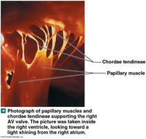
Papillary muscles contract to keep the chordae tendineae taut, preventing AV valve prolapse during ventricular contraction.
Blood Flow Through the Heart
Blood follows a specific path through the heart, ensuring efficient oxygenation and circulation.
Superior/Inferior vena cava → Right atrium → Right ventricle → Pulmonary trunk → Pulmonary arteries → Lungs (capillaries) → Pulmonary veins → Left atrium → Left ventricle → Aorta → Arteries → Body (capillaries) → Veins
Septal Structures
The heart contains septa that separate its chambers.
Interatrial septum: Separates the atria.
Interventricular septum: Separates the ventricles.
Right Atrium
The right atrium receives deoxygenated blood from the body and heart wall.
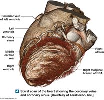
Coronary sinus: Collects blood from the heart wall and delivers it to the right atrium.
Foramen ovale: Fetal structure allowing blood to bypass lungs; becomes fossa ovalis after birth.
Right Ventricle
The right ventricle receives blood from the right atrium and pumps it to the lungs via the pulmonary trunk.
Left Atrium
The left atrium receives oxygenated blood from the pulmonary veins and passes it to the left ventricle.
Left Ventricle
The left ventricle has the thickest wall and pumps oxygenated blood to the aorta and systemic circulation.
Aortic sinuses: Fill and supply coronary circulation.
Valve Function During Cardiac Cycle
Relaxed ventricles: AV valves open, semilunar valves closed.
Contracted ventricles: AV valves closed, semilunar valves open.
Coronary Circulation
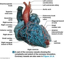
Coronary circulation supplies blood to the heart muscle itself.
Coronary arteries: First branches off the aorta, supply the heart.
Coronary veins: Return blood to the coronary sinus, which empties into the right atrium.
Coronary Artery Disease and Myocardial Infarction
Coronary artery disease (CAD) is a leading cause of death, often resulting from atherosclerosis.
Ischemia: Reduced blood supply to the heart.
Angina: Chest pain due to ischemia.
Myocardial infarction (MI): Heart attack caused by blocked blood vessels, leading to tissue death and scarring.
Cardiac Nodal (Pacemaker) Cells and Conducting System
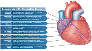
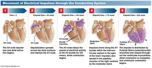
The heart's conducting system coordinates rhythmic contractions via specialized pacemaker cells.
Sinoatrial (SA) node: Initiates heartbeat.
Internodal pathways: Conduct impulses from SA node to AV node.
Atrioventricular (AV) node: Delays impulse, allowing atria to contract before ventricles.
AV bundle, bundle branches, moderator band, Purkinje fibers: Distribute impulse to ventricles for coordinated contraction.
The Cardiac Cycle
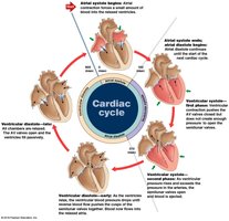
The cardiac cycle describes the sequence of events during a heartbeat, alternating between contraction (systole) and relaxation (diastole).
Systole: Contraction phase, chambers empty.
Diastole: Relaxation phase, chambers fill.
Sequence: 1) Atrial systole, ventricular diastole; 2) Atrial diastole, ventricular systole; 3) Atrial diastole, ventricular diastole.
Abnormal Pacemaker Function
Disorders of pacemaker function can cause arrhythmias.
Bradycardia: Slower-than-normal heart rate.
Tachycardia: Faster-than-normal heart rate.
Autonomic Nervous System and Hormonal Regulation
The heart rate and force of contraction are regulated by the autonomic nervous system and hormones.
Sympathetic nervous system: Norepinephrine (NE) increases heart rate and contraction force via beta receptors.
Parasympathetic nervous system: Acetylcholine (ACh) decreases heart rate via muscarinic receptors.
Cardiac centers in medulla oblongata: Send impulses to adjust heart rate and force.
Summary Table: Heart Valves and Their Locations
Valve | Location | Function |
|---|---|---|
Right AV (Tricuspid) | Between right atrium and right ventricle | Prevents backflow into right atrium |
Left AV (Mitral/Bicuspid) | Between left atrium and left ventricle | Prevents backflow into left atrium |
Pulmonary Semilunar | Between right ventricle and pulmonary trunk | Prevents backflow into right ventricle |
Aortic Semilunar | Between left ventricle and aorta | Prevents backflow into left ventricle |
Summary Table: Cardiac Cycle Phases
Phase | Chamber State | Valve State |
|---|---|---|
Atrial Systole | Atria contract, ventricles relax | AV valves open, semilunar valves closed |
Ventricular Systole | Ventricles contract, atria relax | AV valves closed, semilunar valves open |
Diastole | Both chambers relax | AV valves open, semilunar valves closed |
Key Equations
Cardiac Output (CO): The volume of blood pumped by the heart per minute. Where: = Heart Rate (beats per minute) = Stroke Volume (mL per beat)
Additional info:
Cardiac muscle cells are highly resistant to fatigue due to abundant mitochondria.
Coronary circulation is essential for maintaining heart tissue viability.
Autorhythmicity is a unique property of cardiac muscle, allowing the heart to beat independently of external stimuli.
