 Back
BackThe Heart: Structure, Valves, and Conduction System
Study Guide - Smart Notes
 Tailored notes based on your materials, expanded with key definitions, examples, and context.
The Heart
Overview of Heart Structure
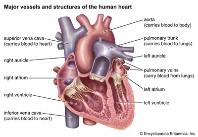
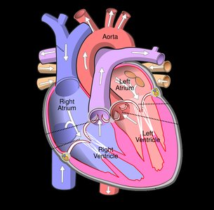
The heart is a muscular organ responsible for pumping blood throughout the body via the circulatory system. It consists of four chambers and several major vessels that ensure unidirectional blood flow.
Right Atrium: Receives deoxygenated blood from the body via the superior and inferior vena cava.
Right Ventricle: Pumps deoxygenated blood to the lungs through the pulmonary trunk.
Left Atrium: Receives oxygenated blood from the lungs via the pulmonary veins.
Left Ventricle: Pumps oxygenated blood to the body through the aorta.
Heart Valves
Heart valves ensure one-way flow of blood and prevent backflow. There are two main types of valves:
Atrioventricular (AV) Valves:
Tricuspid Valve: Located between the right atrium and right ventricle.
Bicuspid (Mitral) Valve: Located between the left atrium and left ventricle.
Both are supported by chordae tendineae, which are attached to papillary muscles to prevent valve prolapse during ventricular contraction.
Semilunar Valves:
Pulmonary Valve: Between the right ventricle and pulmonary trunk.
Aortic Valve: Between the left ventricle and aorta.
These valves are not supported by chordae tendineae.
Valve Disorders:
Regurgitation: Condition where blood flows backward due to improper valve closure.
Stenosis: Condition where a valve does not open completely, restricting blood flow.
Fibrous Pericardium
The fibrous pericardium is a tough outer layer that protects the heart and restricts overfilling or overstretching during blood volume changes.
Cardiac Muscle and Conduction System
The heart is unique in its ability to generate its own action potentials, allowing it to contract rhythmically and independently of external stimuli. This is due to specialized conduction cardiac cells.
Conduction System Pathway:
Sinoatrial (SA) Node: The primary pacemaker, initiates the heartbeat.
Atrioventricular (AV) Node: Receives the impulse from the SA node and delays it to allow atrial contraction.
Atrioventricular (AV) Bundle (Bundle of His): Conducts impulses from the AV node to the ventricles.
Left and Right Bundle Branches: Carry impulses through the interventricular septum.
Purkinje Fibers: Distribute the impulse throughout the ventricular myocardium, causing contraction.
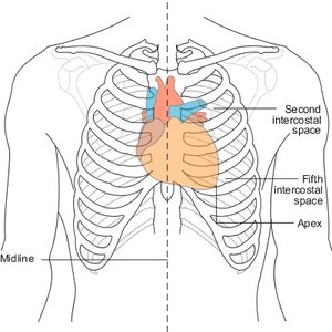
Surface Anatomy of the Heart
Understanding the surface anatomy of the heart is important for clinical assessment and procedures.
Sternal Angle: The second rib attaches here; used as a landmark for counting ribs.
Apex of the Heart: Located at the fifth intercostal space, typically at the midclavicular line.
Key Terms and Definitions
Chordae Tendineae: Tendinous cords that anchor AV valve leaflets to papillary muscles.
Papillary Muscles: Muscles in the ventricles that contract to prevent AV valve prolapse.
Regurgitation: Backflow of blood due to valve incompetence.
Stenosis: Narrowing of a valve opening, impeding blood flow.
Fibrous Pericardium: Outer protective layer of the heart.
Table: Comparison of Heart Valves
Valve | Location | Supported by Chordae Tendineae? |
|---|---|---|
Tricuspid | Right AV (between right atrium and ventricle) | Yes |
Bicuspid (Mitral) | Left AV (between left atrium and ventricle) | Yes |
Pulmonary | Right ventricle to pulmonary trunk | No |
Aortic | Left ventricle to aorta | No |
Additional info:
The heart's ability to generate its own action potentials is termed autorhythmicity.
The sternal angle is a key anatomical landmark for clinicians to locate heart structures during physical examination.
