 Back
BackThe Human Reproductive System: Structure, Development, and Function
Study Guide - Smart Notes
 Tailored notes based on your materials, expanded with key definitions, examples, and context.
Overview of the Reproductive Systems
Introduction to Male and Female Reproductive Anatomy
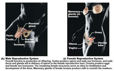
The human reproductive system consists of primary sex organs (gonads), accessory sex organs, and external genitalia. These structures are specialized for the production of gametes, fertilization, and, in females, the nourishment of offspring. Mammary glands, though not sex organs, play a crucial role in postnatal nourishment.
Primary sex organs: Gonads (testes in males, ovaries in females) produce gametes and sex hormones.
Accessory sex organs: Ducts, glands, and external genitalia facilitate gamete transport and fertilization.
Mammary glands: Modified sweat glands in females, responsible for milk production.
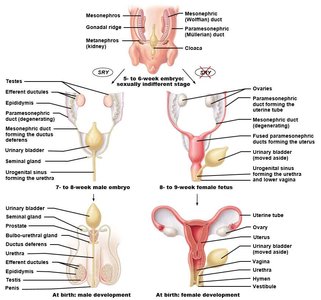
Sexual Differentiation and Homologous Structures
Genetic Sex Determination and Embryonic Development
Genetic sex is determined at conception by the combination of sex chromosomes. Sexual differentiation begins around the 7th to 8th week of embryonic development, primarily regulated by the SRY protein. Homologous structures arise from similar embryonic tissues and differentiate into male or female organs depending on genetic and hormonal signals.
SRY protein: Acts as a genetic switch; its presence initiates male development from the gonadal ridges.
Homologous structures: Internal and external genitalia develop from common embryonic tissues.
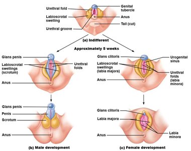
Development of External Genitalia
By the 8th week of development, the external genitalia begin to differentiate. The genital tubercle, labioscrotal swellings, and urethral folds give rise to distinct male and female structures.
Genital tubercle: Forms the glans penis in males and the clitoris in females.
Labioscrotal swelling: Develops into the scrotum in males and labia majora in females.
Urethral folds: Become the spongy urethra in males and labia minora in females.
Testicular descent: Testes descend through the inguinal canal during fetal development.
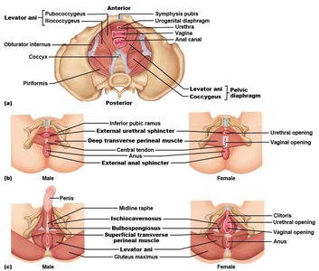
Pelvic Floor and Perineum
Anatomy and Function
The pelvic floor, or pelvic diaphragm, is composed of muscles that support the pelvic organs. The perineum is the region inferior to the pelvic floor, bounded by the pubic symphysis, ischial tuberosities, and coccyx. It contains the urogenital and anal sphincters, as well as muscles involved in sexual function.
Pelvic diaphragm: Includes the levator ani and coccygeus muscles.
Perineum: Contains the urogenital diaphragm, external urethral and anal sphincters, bulbospongiosus, and ischiocavernosus muscles.
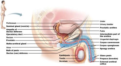
Male Reproductive System
Gross Anatomy
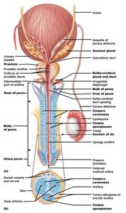
The male reproductive system includes the testes, duct system, accessory glands, and external genitalia. The testes produce sperm, which travel through a series of ducts and are mixed with glandular secretions to form semen.
Testes: Site of gamete (sperm) production.
Duct system: Epididymis, ductus deferens, ejaculatory duct, and urethra transport sperm.
Accessory glands: Seminal vesicles, prostate, and bulbourethral glands contribute fluids to semen.
External genitalia: Penis and scrotum.
Testes and Epididymis
The testes are enclosed by the tunica vaginalis and tunica albuginea, and are divided into lobules containing seminiferous tubules. Sperm produced in the seminiferous tubules travel through straight tubules and the rete testis to the epididymis, where they mature and are stored.
Tunica vaginalis: Embryonic remnant of peritoneum surrounding the testis.
Tunica albuginea: Dense connective tissue capsule.
Seminiferous tubules: Site of spermatogenesis.
Epididymis: Coiled duct where sperm mature and are stored.
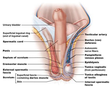
Spermatic Cord and Scrotum
The spermatic cord is a connective tissue sheath containing ducts, nerves, blood vessels, and muscles. The cremaster muscle elevates or lowers the testis, while the dartos muscle wrinkles or smooths the scrotal skin to regulate temperature.
Spermatic cord: Extends from the abdomen to the scrotum, passing through the inguinal canal.
Cremaster muscle: Raises and lowers the testis in response to temperature changes.
Dartos muscle: Adjusts the surface area of the scrotum.
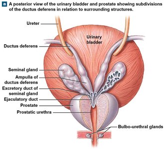
Duct System
The duct system transports sperm from the testes to the urethra. Efferent ductules connect the rete testis to the epididymis, which leads to the ductus deferens. The ductus deferens joins the seminal vesicle duct to form the ejaculatory duct, which empties into the urethra.
Efferent ductules: Connect rete testis to epididymis.
Epididymis: Site of sperm maturation and storage.
Ductus deferens: Transports sperm during ejaculation; expands into the ampulla before joining the ejaculatory duct.
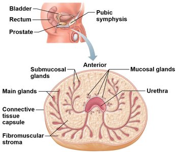
Accessory Glands
Accessory glands produce secretions that combine with sperm to form semen. The seminal vesicles contribute most of the semen volume, the prostate surrounds the urethra and secretes enzymes, and the bulbourethral glands secrete mucus for lubrication and pH neutralization.
Seminal vesicles: Produce 60% of semen volume; ducts empty into ejaculatory duct.
Prostate: Surrounds urethra; secretes enzymes that clot and liquefy semen.
Bulbourethral glands: Inferior to prostate; secrete mucus for lubrication.
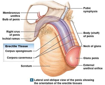
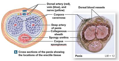
External Genitalia
The penis consists of the root, body, and glans penis. The glans is covered by the prepuce, which may be removed during circumcision. Internally, the penis contains three erectile bodies: one corpus spongiosum (ventral, surrounding the urethra) and two corpora cavernosa (dorsal).
Corpus spongiosum: Surrounds the urethra and forms the glans penis.
Corpora cavernosa: Paired dorsal erectile bodies.
Female Reproductive System
Gross Anatomy
The female reproductive system includes the ovaries, duct system (uterine tubes, uterus, vagina), and external genitalia (vulva). Mammary glands are not directly involved in reproduction but are hormonally regulated and essential for infant nourishment.
Ovaries: Site of oocyte production and hormone secretion.
Duct system: Uterine tubes, uterus, and vagina transport gametes and support fertilization and fetal development.
External genitalia: Vulva, including mons pubis, labia majora, labia minora, vestibule, and clitoris.
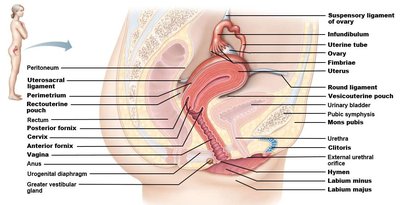
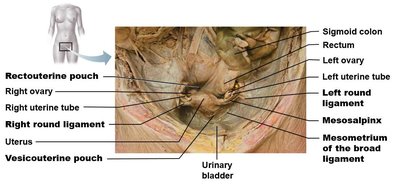
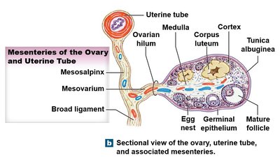
Ligaments and Uterine Tubes
Ligaments anchor the ovaries and uterus within the pelvic cavity. The broad ligament is a fold of peritoneum, while the suspensory and ovarian ligaments provide additional support. The uterine tubes (fallopian tubes) capture oocytes and transport them to the uterus.
Broad ligament: Anchors uterine tubes and uterus.
Suspensory ligament: Attaches ovary to pelvic wall; contains vessels and nerves.
Ovarian ligament: Anchors ovary to uterus.
Uterine tube regions: Fimbriae, infundibulum, ampulla, isthmus.
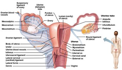
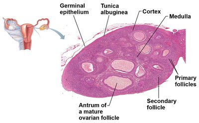
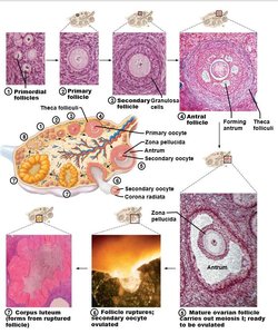
Ovarian Structure and Follicular Development
The ovaries are retroperitoneal organs located near the sacroiliac joint. Each ovary is covered by a tunica albuginea and contains a cortex (with follicles) and medulla (with vessels and nerves). Oocytes develop within follicles, which progress through several stages before ovulation.
Follicular stages: Primordial, primary, secondary, antral, mature (tertiary) follicles.
Ovulation: Release of oocyte from mature follicle.
Corpus luteum: Temporary endocrine structure formed after ovulation.
Corpus albicans: Degenerated corpus luteum.
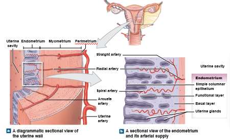
Uterus and Uterine Wall
The uterus is a muscular organ superior to the bladder and anterior to the rectum. It consists of the body, fundus, and cervix. The uterine wall has three layers: perimetrium (serous), myometrium (muscular), and endometrium (mucosal). The endometrium has two sub-layers: the basalis (regenerative) and functionalis (shed during menstruation).
Cervix: Neck of the uterus; closed during pregnancy, dilates during labor.
Endometrium: Site of implantation and menstruation.
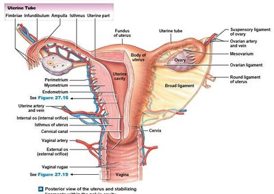
Vagina
The vagina is a muscular canal extending from the cervix to the vaginal orifice. Its mucosal lining is folded into rugae, and the superior fornix surrounds the cervix. The hymen is an incomplete diaphragm near the vaginal orifice.
Fornix: Recess around the cervix.
Hymen: Thin membrane partially covering the vaginal opening.
Anatomical Changes During the Menstrual Cycle
The menstrual cycle involves cyclical changes in the endometrium. The menstrual phase involves shedding of the functionalis layer, the proliferative phase rebuilds this layer, and the secretory phase enriches it in preparation for possible implantation.
Menstrual phase: Shedding of the functional layer of the endometrium.
Proliferative phase: Rebuilding of the functional layer (pre-ovulation).
Secretory phase: Enrichment of the functional layer (post-ovulation).
External Genitalia (Vulva)
The vulva includes all external female genital structures. It is distinct from the vagina and includes the mons pubis, labia majora, labia minora, vestibule (containing urethral and vaginal openings), and clitoris.
Mons pubis: Fatty area overlying the pubic symphysis.
Labia majora: Outer folds covering the labia minora.
Labia minora: Inner folds enclosing the vestibule.
Clitoris: Erectile tissue anterior to the vestibule.
Breast and Mammary Glands
The breasts extend from the 2nd to 6th rib and from the sternum to the mid-axillary line. Each breast contains 15-25 lobes separated by suspensory ligaments and adipose tissue. Mammary glands are compound alveolar glands that produce milk, which is delivered through lactiferous ducts and sinuses to the nipple.
Mammary glands: Specialized for milk production.
Nipple: Contains openings of lactiferous ducts.
Summary Table: Homologous Structures of Male and Female Reproductive Systems
Embryonic Structure | Male Derivative | Female Derivative |
|---|---|---|
Genital tubercle | Glans penis | Clitoris |
Labioscrotal swelling | Scrotum | Labia majora |
Urethral folds | Spongy urethra | Labia minora |
Gonadal ridge | Testis | Ovary |
