 Back
BackThe Human Reproductive System: Structure, Function, and Regulation
Study Guide - Smart Notes
 Tailored notes based on your materials, expanded with key definitions, examples, and context.
The Reproductive System: Overview
Introduction to the Reproductive System
The reproductive system is unique among organ systems because it is not essential for the survival of the individual, but it is crucial for the continuation of the species. It interacts with other body systems through hormone secretion and physiological effects. The primary organs, called gonads, produce gametes and hormones, while ducts transport gametes, and accessory glands secrete fluids that support reproductive function. External genitalia are the visible structures associated with reproduction.
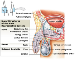
Structures of the Male Reproductive System
Major Structures and Functions
The male reproductive system is specialized for the production, maturation, and delivery of spermatozoa. It includes the testes (gonads), a series of ducts, accessory glands, and external genitalia.
Testes: Produce sperm and secrete androgens (male sex hormones).
Ducts: Transport sperm from the testes to the exterior (epididymis, ductus deferens, ejaculatory duct, urethra).
Accessory glands: Seminal vesicles, prostate gland, and bulbourethral glands add secretions to semen.
External genitalia: Penis (delivers sperm) and scrotum (houses testes).
Pathway of Spermatozoa
Sperm are produced in the testes and travel through a series of ducts before exiting the body:
Testis
Epididymis
Ductus deferens (vas deferens)
Ejaculatory duct
Urethra
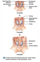
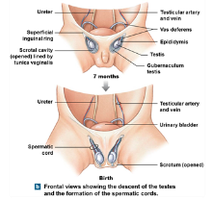
Descent of the Testes
During fetal development, the testes descend from their origin near the kidneys into the scrotum. This process is guided by the gubernaculum testis and is essential for normal sperm production, which requires a temperature lower than core body temperature.
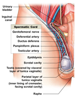
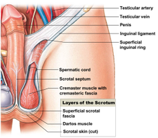
The Spermatic Cord
The spermatic cord contains the ductus deferens, blood vessels, nerves, and lymphatics that supply the testes. It passes through the inguinal canal and is essential for testicular function and support.
Scrotum and Associated Muscles
The scrotum is a pouch of skin and muscle that houses the testes. Two muscles regulate testicular temperature:
Dartos muscle: Smooth muscle that wrinkles the scrotal skin.
Cremaster muscle: Skeletal muscle that raises or lowers the testes.

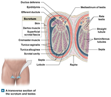
Internal Structure of the Testes
The testes are divided into lobules containing seminiferous tubules, where spermatogenesis occurs. The tubules are surrounded by connective tissue and contain various cell types, including spermatogenic cells and Sertoli (nurse) cells.
Spermatogenesis and Sperm Structure
Spermatogenesis
Spermatogenesis is the process of sperm production, occurring in the seminiferous tubules. It involves three main phases:
Mitosis: Spermatogonia divide to produce primary spermatocytes.
Meiosis: Primary spermatocytes undergo meiosis to form spermatids.
Spermiogenesis: Spermatids mature into spermatozoa (sperm).
This process takes about nine weeks from spermatogonial division to the release of mature sperm into the lumen (spermiation).
Role of Sertoli (Nurse) Cells
Sertoli cells support and regulate spermatogenesis by:
Maintaining the blood-testis barrier
Supporting mitosis and meiosis
Secreting inhibin, androgen-binding protein (ABP), and Müllerian-inhibiting factor (MIF)
Anatomy of a Spermatozoon
A mature sperm cell consists of:
Head: Contains the nucleus and acrosome (enzymes for fertilization)
Neck: Connects head to middle piece
Middle piece: Packed with mitochondria for energy
Tail: A flagellum for motility
Male Reproductive Ducts and Glands
Epididymis
The epididymis is a coiled tube where sperm mature and are stored. It monitors and adjusts the fluid from the seminiferous tubules, recycles damaged sperm, and stores spermatozoa.
Ductus Deferens (Vas Deferens)
This muscular tube transports sperm from the epididymis to the ejaculatory duct. It can store sperm for several months in a low metabolic state.
Ejaculatory Duct and Urethra
The ejaculatory duct is a short passage that joins the ductus deferens with the seminal gland duct and empties into the urethra, which serves both urinary and reproductive functions in males.
Accessory Glands
Accessory glands produce the bulk of semen and provide nutrients, buffers, and enzymes for sperm function. The main glands are:
Seminal vesicles: Produce 60% of semen volume, rich in fructose and prostaglandins.
Prostate gland: Secretes a milky, alkaline fluid with enzymes and antibiotic seminalplasmin.
Bulbourethral glands: Secrete alkaline mucus for lubrication and neutralization of urinary acids.
Hormonal Regulation of Male Reproduction
Key Hormones
Gonadotropin-releasing hormone (GnRH): From the hypothalamus, stimulates the anterior pituitary to release FSH and LH.
Follicle-stimulating hormone (FSH): Stimulates spermatogenesis and Sertoli cell function.
Luteinizing hormone (LH): Stimulates testosterone production by interstitial (Leydig) cells.
Testosterone: Promotes development of male secondary sex characteristics and supports spermatogenesis.
Inhibin: Produced by Sertoli cells, inhibits FSH release to regulate sperm production.
Negative feedback mechanisms involving testosterone and inhibin maintain hormonal balance and regulate sperm production.
Summary Table: Major Structures of the Male Reproductive System
Structure | Function |
|---|---|
Testis | Produces sperm and testosterone |
Epididymis | Sperm maturation and storage |
Ductus deferens | Transports and stores sperm |
Seminal vesicle | Secretes fructose-rich fluid |
Prostate gland | Secretes alkaline fluid and enzymes |
Bulbourethral gland | Secretes mucus for lubrication |
Penis | Delivers sperm to female tract |
Scrotum | Houses and cools testes |
Additional info: This summary covers the male reproductive system. The female reproductive system, reproductive cycles, and sexual function are also essential topics in human anatomy and physiology and are covered in subsequent sections of the source material.
