 Back
BackThe Human Skeleton: Axial and Appendicular Divisions, Skull Anatomy, and Bone Markings
Study Guide - Smart Notes
 Tailored notes based on your materials, expanded with key definitions, examples, and context.
The Human Skeleton
Overview of the Skeletal System
The human skeleton is a structural framework composed of bones, cartilages, joints, and ligaments. It is divided into two major divisions: the axial skeleton and the appendicular skeleton. The skeleton accounts for approximately 20% of body mass and serves to support, protect, and facilitate movement.
Axial skeleton: Consists of the skull, vertebral column, and thoracic cage. Forms the longitudinal axis of the body.
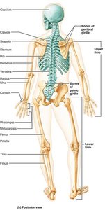
Appendicular skeleton: Includes the bones of the limbs and girdles (pectoral and pelvic) that attach them to the axial skeleton.
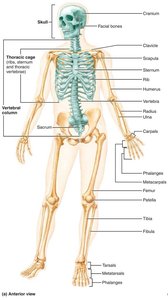
The Axial Skeleton
Skull: Structure and Function
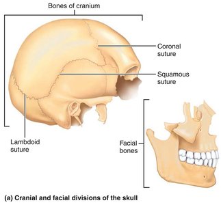
The skull is formed by two sets of bones: cranial bones and facial bones. Cranial bones enclose the brain and provide attachment sites for head and neck muscles, while facial bones form the framework of the face and house cavities for sensory organs.
Cranial bones: Enclose and protect the brain; provide muscle attachment sites.
Facial bones: Form the face; contain cavities for sight, taste, and smell; provide openings for air and food passage; secure teeth; anchor facial muscles.
Sutures: Immovable joints connecting skull bones, with a serrated, saw-tooth appearance.
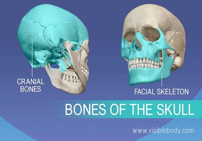
Comparison of Cranium and Facial Skeleton Functions
Cranium: Protection of the brain; attachment for head and neck muscles.
Facial skeleton: Framework for the face; cavities for sensory organs; muscle attachment for facial expression.
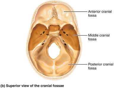
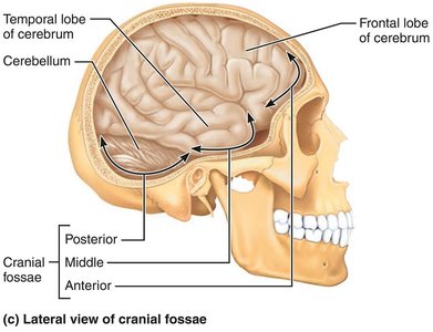
Skull Geography and Cavities
The cranium is divided into a vault (calvaria) and a base. The cranial base is internally divided into three fossae: anterior, middle, and posterior, which house different regions of the brain. The skull also contains other cavities such as the middle and internal ear, nasal cavity, orbits, and sinuses.
Cranial vault: Forms the superior, lateral, and posterior portion of the skull.
Cranial base: Forms the inferior aspect; divided into fossae.
Openings: Foramina, canals, and fissures provide passageways for nerves and blood vessels.
Skull Bone Markings
Foramina: Openings for blood vessels and nerves.
Processes: Projections for muscle attachment.
Sutures: Immovable joints connecting skull bones.
Cranial Bones
The cranium comprises eight cranial bones: frontal, parietal (left and right), occipital, temporal (left and right), sphenoid, and ethmoid. 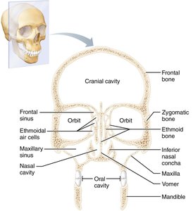
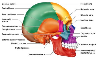
Frontal Bone
The frontal bone forms the anterior portion of the cranium, including the forehead and superior wall of the orbits.
Squamous region: Forehead.
Supraorbital margin: Area underneath eyebrows.
Supraorbital foramen: Passage for artery and nerve.
Glabella: Area between orbits.
Frontal sinuses: Located lateral to glabella.
Parietal Bones and Sutures
Two large parietal bones form most of the superior and lateral aspects of the cranial vault. Four major sutures mark their articulations: coronal, sagittal, lambdoid, and squamous.
Occipital Bone
The occipital bone forms the posterior wall and base of the skull.
Foramen magnum: Passage for the spinal cord.
Occipital condyles: Articulate with the first vertebra.
External occipital protuberance and crest: Sites for ligament and muscle attachment.
Nuchal lines: Attachment for neck and back muscles.
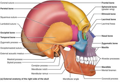
Temporal Bones
Paired temporal bones make up the inferolateral aspects of the skull and parts of the cranial base.
Squamous part: Zygomatic process forms zygomatic arch; mandibular fossa forms part of temporomandibular joint.
Tympanic part: Surrounds external acoustic meatus.
Petrous part: Houses middle and internal ear cavities; contains several foramina for nerves and arteries.
Mastoid and styloid processes: Muscle attachment sites.

Sphenoid Bone
The sphenoid bone is a complex, bat-shaped bone that articulates with all other cranial bones.
Sphenoidal sinuses: Within the body of the sphenoid.
Sella turcica: Encloses the pituitary gland.
Processes: Greater wings, lesser wings, pterygoid processes.
Foramina: Optic canals, superior orbital fissure, foramen rotundum, foramen ovale, foramen spinosum.
Ethmoid Bone
The ethmoid bone is the deepest skull bone, forming the roof of the nasal cavity and part of the anterior cranial fossa.
Cribriform plates: Roof of nasal cavity.
Crista galli: Attachment for dura mater.
Perpendicular plate: Superior part of nasal septum.
Ethmoidal labyrinths: Contain ethmoidal air cells.
Orbital plates: Medial wall of orbits.
Sutural Bones
Sutural bones are tiny, irregularly shaped bones that appear within sutures. Their significance is unknown and not everyone has them.
Facial Bones
The facial skeleton is made up of 14 bones, 12 of which are paired.
Mandible: Largest, strongest bone of the face.
Maxillary bones: Form upper jaw and central facial skeleton.
Zygomatic bones: Form cheekbones.
Nasal bones: Form bridge of nose.
Lacrimal bones: Form medial walls of orbits.
Palatine bones: Complete hard palate and form part of nasal cavity and orbits.
Vomer: Forms part of nasal septum.
Inferior nasal conchae: Form part of lateral walls of nasal cavity.
Bony Boundaries of the Orbits and Nasal Cavity
Orbits: Roof (frontal, sphenoid), lateral wall (zygomatic, sphenoid), floor (maxilla, zygomatic, palatine), medial wall (ethmoid, lacrimal, maxilla, sphenoid).
Nasal cavity: Roof (nasal, frontal, ethmoid, sphenoid), floor (maxilla, palatine), lateral walls (maxilla, ethmoid, inferior nasal conchae, palatine), medial wall (ethmoid, vomer).
Paranasal sinuses: Frontal, ethmoidal, sphenoidal, maxillary sinuses.
Paranasal Sinuses
Paranasal sinuses are hollow portions of bones surrounding the nasal cavity.
Functions: Lighten the skull; give resonance and amplification to the voice.
The Fetal Skull
The fetal skull is large compared to the infant's total body length and has more bones than the adult skull.
Fontanelles: Unossified remnants of fibrous membranes; ease birth and allow brain growth.
Conversion: Fontanelles convert to bone within 24 months after birth.
The Vertebral Column
General Characteristics
The vertebral column extends from the skull to the pelvis and functions to transmit weight, protect the spinal cord, and provide attachment points for ribs and muscles.
Regions: Cervical (7), thoracic (12), lumbar (5), sacrum (5 fused), coccyx (4 fused).
Curvatures: Cervical and lumbar (convex anteriorly), thoracic and sacral (concave anteriorly).
Functions of Spinal Curvatures and Intervertebral Discs
Spinal curvatures: Increase resilience and flexibility; help maintain balance and posture.
Intervertebral discs: Cushion vertebrae; allow bending and twisting; contribute to shock absorption.
Abnormal Spinal Curvatures
Scoliosis: Abnormal lateral rotation; may lead to breathing difficulties.
Kyphosis: Abnormal dorsal thoracic curvature; common in osteoporosis.
Lordosis: Accentuated lumbar curvature; seen in pregnancy and obesity.
Structure of a Typical Vertebra
Body: Main weight-bearing structure.
Vertebral arch: Formed by pedicles and laminae.
Processes: Spinous, transverse, superior and inferior articular processes.
Regional Features of Vertebrae
Cervical: Smallest; transverse foramina; bifid spinous processes.
Thoracic: Articular facets for ribs; heart-shaped body; long, slender spinous processes.
Lumbar: Largest; kidney-shaped body; short, blunt spinous processes.
The Thoracic Cage
Bones of the Thoracic Cage
The thoracic cage forms a protective cage for major organs and is composed of the sternum, ribs, and thoracic vertebrae.
Sternum: Manubrium, body, xiphoid process.
Ribs: True ribs (1-7), false ribs (8-12), floating ribs (11-12).
Functions: Protection, support, respiration.
True vs. False Ribs
True ribs: Attach directly to the sternum via their own costal cartilage.
False ribs: Attach indirectly or not at all; floating ribs do not attach to the sternum.
The Appendicular Skeleton
Pectoral (Shoulder) Girdle
The pectoral girdle is composed of the clavicle and scapula, allowing for free movement of the upper limb.
Clavicle: Collarbone; fractures anteriorly to protect subclavian artery.
Scapula: Shoulder blade; triangular with three borders; prominent spine on posterior surface.
Bones of the Upper Limb
Humerus: Arm bone; articulates with scapula, radius, and ulna.
Ulna: Main responsibility for forming elbow joint; olecranon and coronoid processes.
Radius: Major contribution to wrist joint; radial styloid process.
Hand: Carpals (wrist), metacarpals (palm), phalanges (fingers).
Pelvic Girdle
Hip bones: Ilium, ischium, pubis; fused to form acetabulum.
Functions: Supports upper body weight; protects organs.
Female pelvis: Wider, shallower, lighter, rounder for childbearing.
Bones of the Lower Limb
Femur: Thigh bone; largest, longest, strongest bone.
Patella: Knee cap; protects knee joint.
Tibia: Medial, thicker; bears weight.
Fibula: Lateral, finer; does not bear weight.
Foot: Tarsals (ankle), metatarsals (sole), phalanges (toes).
Arches: Medial and lateral longitudinal, transverse; maintained by bone shapes, ligaments, tendons.
Joints
Classification of Joints
Joints are classified functionally and structurally.
Functional: Synarthroses (immovable), amphiarthroses (slightly movable), diarthroses (freely movable).
Structural: Fibrous, cartilaginous, synovial.
Fibrous Joints
Suture: Only in skull; immovable.
Syndesmosis: Bones connected by ligaments; movement depends on fiber length.
Gomphosis: Peg-in-socket; only example is tooth in alveolar socket.
Cartilaginous Joints
Synchondrosis: Hyaline cartilage unites bones; immovable.
Symphysis: Fibrocartilage unites bones; slightly movable (e.g., pubic symphysis, intervertebral joints).
Synovial Joints
Articular cartilage: Covers ends of bones.
Joint capsule: Encloses joint surfaces; fibrous layer and synovial membrane.
Joint cavity: Filled with synovial fluid.
Ligaments: Reinforce the joint.
Structures Associated with Synovial Joints
Bursae: Flattened fibrous sacs lined with synovial membrane; filled with synovial fluid.
Tendon sheath: Elongated bursa wrapping around a tendon.
Factors Affecting Synovial Joint Stability
Articular surface shape
Number and positioning of ligaments
Muscle tone
Types of Synovial Joints Based on Shape
Plane, hinge, pivot, condyloid, saddle, ball-and-socket
Inflammatory Conditions Associated with Joints
Bursitis: Inflammation of a bursa.
Tendonitis: Inflammation of tendon sheaths.
Arthritis: Inflammatory or degenerative diseases of joints; includes osteoarthritis and rheumatoid arthritis.
Clinical and Bone Health Considerations
Bone Imaging Techniques
X-rays: Detailed images of bone and soft tissues.
MRI: Cross-sectional images for complex fractures.
CT scans: Detailed bone structure analysis.
Prevention and Treatment of Bone Disorders
Medications: Bisphosphonates, SERMs, hormone therapy.
Prevention: Calcium, vitamin D, weight-bearing exercise, avoiding smoking and excessive alcohol.
Risk factors: Age, genetics, lifestyle, decreased estrogen in postmenopausal women.
Role of Hormones in Bone Health
Estrogen: Promotes bone deposition; decreased levels contribute to bone loss.
Testosterone: Promotes bone deposition and density.
Parathyroid hormone (PTH): Regulates calcium; stimulates bone resorption.
Exercise Guidelines for Strong Bones
Weight-bearing exercises: Walking, running, dancing, weightlifting.
Resistance training: Strengthens muscles and bones.
Flexibility exercises: Improve range of motion.
Screening for Bone Health
Bone density screening: DEXA scan.
Risk assessments: Age, genetics, lifestyle.
Early detection: Allows timely intervention.
Role of Rehabilitation in Bone Healing
Occupational therapy: Regains independence in daily activities.
Physical therapy: Improves strength, flexibility, balance.
Post-fracture rehabilitation: Restores function and mobility.
