 Back
BackThe Human Skeleton: Axial and Appendicular Divisions, Skull Anatomy, and Bone Markings
Study Guide - Smart Notes
 Tailored notes based on your materials, expanded with key definitions, examples, and context.
The Human Skeleton
Overview and Divisions
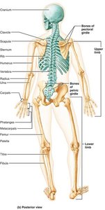
The human skeleton is a structural framework composed of bones, cartilages, joints, and ligaments. It accounts for approximately 20% of body mass and is divided into two major regions: the axial skeleton and the appendicular skeleton.
Axial Skeleton: Consists of 80 bones, including the skull, vertebral column, and thoracic cage. It forms the longitudinal axis of the body, supports the head, neck, and trunk, and protects the brain, spinal cord, and thoracic organs.
Appendicular Skeleton: Comprises the bones of the limbs and girdles, facilitating movement and interaction with the environment.
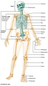
The Axial Skeleton
Skull: Structure and Function
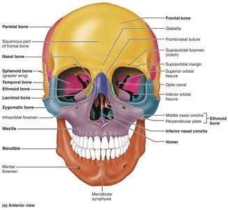
The skull is formed by two sets of bones: cranial bones and facial bones.
Cranial Bones: Enclose the brain in the cranial cavity and provide attachment sites for head and neck muscles.
Facial Bones: Form the framework of the face, contain cavities for special sense organs, provide openings for air and food passage, secure teeth, and anchor facial muscles used for expression.
Sutures: Immovable joints connecting skull bones, with a serrated, saw-tooth appearance.
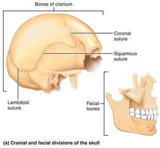
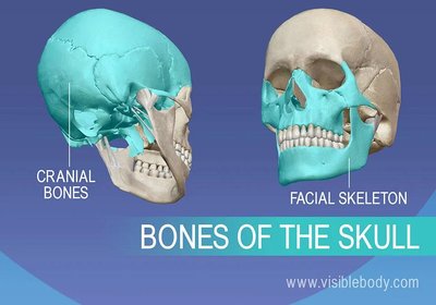
Comparison of Cranium and Facial Skeleton Functions
Cranium: Protects the brain and provides attachment sites for muscles.
Facial Skeleton: Forms the face, houses sensory organ cavities, and provides muscle attachment points.
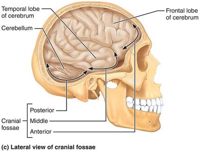
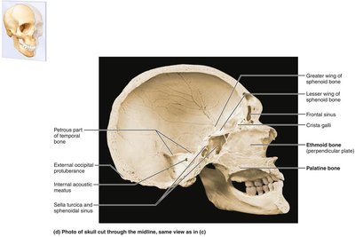
Skull Geography and Cavities
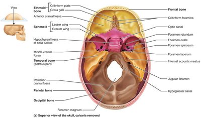
The cranium is divided into a vault (calvaria) and a base. The cranial base is internally divided into three fossae: anterior, middle, and posterior, which house the brain. The skull also contains other cavities, including the middle and internal ear, nasal cavity, orbits, and sinuses. 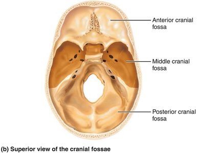
Skull Openings and Markings
Foramina: Openings for blood vessels and nerves.
Processes: Projections for muscle attachment.
Sutures: Immovable joints connecting skull bones.
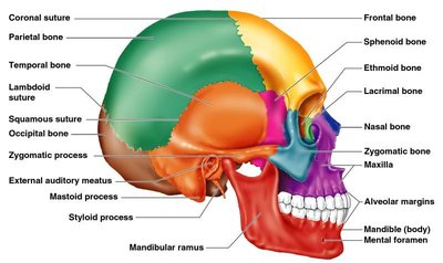
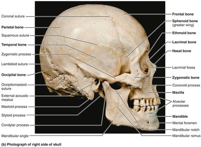
Cranial Bones: Identification and Markings
The cranium comprises eight bones: frontal, parietal (left and right), occipital, temporal (left and right), sphenoid, and ethmoid. 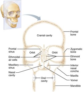

Frontal Bone
Location: Forms the anterior portion of the cranium, including the forehead.
Markings: Supraorbital margin, supraorbital foramen, glabella, and frontal sinuses.
Parietal Bones and Sutures
Location: Form most of the superior and lateral aspects of the cranial vault.
Sutures: Coronal, sagittal, lambdoid, and squamous sutures.

Occipital Bone
Location: Forms the posterior wall and posterior cranial fossa.
Markings: Foramen magnum, occipital condyles, hypoglossal canal, external occipital protuberance, external occipital crest, superior and inferior nuchal lines.

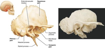
Temporal Bones
Location: Inferolateral aspects of the skull and parts of the cranial base.
Parts: Squamous, tympanic, petrous.
Markings: Zygomatic process, mandibular fossa, external acoustic meatus, mastoid and styloid processes, carotid canal, jugular foramen, foramen lacerum, internal acoustic meatus.

Sphenoid Bone
Location: Keystone bone articulating with all other cranial bones.
Markings: Sella turcica, hypophyseal fossa, greater and lesser wings, pterygoid processes, optic canals, superior orbital fissure, foramen rotundum, foramen ovale, foramen spinosum.
Ethmoid Bone
Location: Deepest skull bone, forms roof of nasal cavity and floor of anterior cranial fossa.
Markings: Cribriform plates, crista galli, perpendicular plate, ethmoidal labyrinths, superior and middle nasal conchae, orbital plates.
Sutural Bones
Definition: Tiny, irregularly shaped bones appearing within sutures; not present in all individuals.
Facial Bones: Identification and Markings
The facial skeleton is made up of 14 bones, 12 of which are paired.
Mandible: Largest, strongest bone of the face; contains alveolar process, mandibular symphysis, coronoid and condylar processes, mandibular and mental foramina.
Maxillae: Form upper jaw and central facial skeleton; contain alveolar processes, palatine process, frontal process, zygomatic processes, maxillary sinuses.
Zygomatic Bones: Form cheekbones and inferolateral margins of orbits.
Nasal Bones: Form bridge of nose.
Lacrimal Bones: Form medial walls of orbits; contain lacrimal fossa.
Palatine Bones: L-shaped bones forming posterior hard palate and part of nasal cavity and orbits.
Vomer: Plow-shaped bone forming part of nasal septum.
Inferior Nasal Conchae: Form part of lateral walls of nasal cavity.
Bony Boundaries of the Orbits and Nasal Cavity
Orbits: Roof (frontal, sphenoid), lateral wall (zygomatic, sphenoid), floor (maxilla, zygomatic, palatine), medial wall (ethmoid, lacrimal, maxilla, sphenoid).
Nasal Cavity: Roof (nasal, frontal, ethmoid, sphenoid), floor (maxilla, palatine), lateral walls (maxilla, ethmoid, inferior nasal conchae, palatine), medial wall (ethmoid, vomer).
Paranasal Sinuses: Frontal, ethmoidal, sphenoidal, maxillary sinuses.
The Vertebral Column
Structure and Regions
The vertebral column extends from the skull to the pelvis and functions to transmit weight, protect the spinal cord, and provide attachment points for ribs and muscles.
Regions: Cervical (7), thoracic (12), lumbar (5), sacrum (5 fused), coccyx (4 fused).
Curvatures: Cervical and lumbar (convex anteriorly), thoracic and sacral (concave anteriorly).
Functions of Spinal Curvatures and Intervertebral Discs
Spinal Curvatures: Increase resilience and flexibility, help maintain balance and posture.
Intervertebral Discs: Cushion vertebrae, allow for bending and twisting, contribute to shock absorption.
Structure of a Typical Vertebra
Body: Main weight-bearing structure.
Vertebral Arch: Formed by pedicles and laminae.
Processes: Spinous, transverse, superior and inferior articular processes.
Regional Features of Vertebrae
Cervical: Smallest, transverse foramina, bifid spinous processes.
Thoracic: Articular facets for ribs, heart-shaped body, long spinous processes.
Lumbar: Largest, kidney-shaped body, short blunt spinous processes.
The Thoracic Cage
Bones and Functions
The thoracic cage forms a protective cage for major organs and is composed of the sternum, ribs, and thoracic vertebrae.
Sternum: Manubrium, body, xiphoid process.
Ribs: True ribs (1-7), false ribs (8-12), floating ribs (11-12).
Functions: Protection, support, respiration.
True vs. False Ribs
True Ribs: Attach directly to the sternum via their own costal cartilage.
False Ribs: Attach indirectly or not at all; floating ribs do not attach to the sternum.
The Appendicular Skeleton
Pectoral Girdle
Clavicle: Collarbone; medial end joins sternum, distal end meets scapula.
Scapula: Shoulder blade; triangular with three borders, prominent spine on posterior surface.
Bones of the Upper Limb
Humerus: Arm bone; articulates with scapula, radius, and ulna.
Ulna: Main responsibility for forming elbow joint; olecranon and coronoid processes.
Radius: Major contribution to wrist joint; radial styloid process.
Hand: Carpals (wrist), metacarpals (palm), phalanges (fingers).
Pelvic Girdle
Hip Bones: Ilium, ischium, pubis; fused to form acetabulum.
Functions: Supports upper body weight, protects organs.
Bones of the Lower Limb
Femur: Thigh bone; largest, longest, strongest bone.
Patella: Knee cap; protects knee joint.
Tibia: Medial, weight-bearing bone.
Fibula: Lateral, non-weight-bearing bone.
Foot: Tarsals (ankle), metatarsals (sole), phalanges (toes).
Joints
Classification and Structure
Joints are articulations of bones, classified functionally and structurally.
Functional Classification: Synarthroses (immovable), amphiarthroses (slightly movable), diarthroses (freely movable).
Structural Classification: Fibrous, cartilaginous, synovial.
Fibrous Joints
Suture: Only found in skull; immovable.
Syndesmosis: Bones connected by ligaments; movement depends on fiber length.
Gomphosis: Peg-in-socket joint; example is tooth in alveolar socket.
Cartilaginous Joints
Synchondrosis: Hyaline cartilage unites bones; immovable.
Symphysis: Fibrocartilage unites bones; examples include pubic symphysis and intervertebral joints.
Synovial Joints
Features: Articular cartilage, joint cavity filled with synovial fluid, fibrous articular capsule, synovial membrane, ligaments.
Bursae: Flattened fibrous sacs lined with synovial membrane.
Tendon Sheath: Elongated bursa wrapping around a tendon.
Factors Affecting Synovial Joint Stability
Shape of articular surfaces
Number and positioning of ligaments
Muscle tone
Inflammatory Conditions Associated with Joints
Bursitis: Inflammation of a bursa.
Tendonitis: Inflammation of tendon sheaths.
Arthritis: Over 100 types; includes osteoarthritis (aging-related) and rheumatoid arthritis (autoimmune).
Clinical and Bone Health Considerations
Bone Imaging Techniques
X-rays: Detailed images for diagnosing fractures and abnormalities.
MRI: Cross-sectional images for complex fractures.
CT Scans: Detailed bone structure analysis.
Prevention and Treatment of Bone Disorders
Medications: Bisphosphonates, SERMs, hormone therapy.
Prevention: Calcium and vitamin D intake, weight-bearing exercise, avoiding smoking and excessive alcohol.
Risk Factors: Age, genetics, lifestyle, decreased estrogen in postmenopausal women.
Role of Hormones in Bone Health
Estrogen: Promotes bone deposition, inhibits resorption.
Testosterone: Increases bone density.
Parathyroid Hormone (PTH): Regulates calcium levels, stimulates bone resorption.
Exercise Guidelines for Strong Bones
Weight-bearing exercises
Resistance training
Flexibility exercises
Screening and Rehabilitation
Bone Density Screening: DEXA scan for osteoporosis risk.
Rehabilitation: Physical and occupational therapy for post-fracture recovery.
Additional info:
Tables and diagrams referenced in the notes are described in text and visually supported by included images.
All equations and formulas are not applicable for this anatomical content.
