 Back
BackThe Human Skeleton: Structure and Function
Study Guide - Smart Notes
 Tailored notes based on your materials, expanded with key definitions, examples, and context.
The Human Skeleton
Overview of the Skeletal System
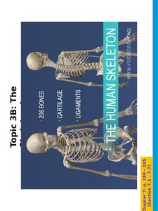
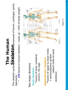
The human skeleton is a complex framework composed of bones, cartilage, and ligaments. It provides structural support, protects vital organs, and facilitates movement. The adult human skeleton consists of 206 bones and accounts for approximately 20% of total body weight.
Bones: Rigid organs that form the skeleton's structure.
Cartilage: Flexible connective tissue found in joints, ear, nose, and other areas.
Ligaments: Strong bands of connective tissue that connect bones at joints.
Divisions of the Skeleton
The skeleton is divided into two major parts:
Axial Skeleton: Includes the skull, vertebral column, and thoracic cage. It forms the central axis of the body.
Appendicular Skeleton: Comprises the bones of the upper and lower limbs, as well as the pectoral and pelvic girdles, which attach the limbs to the axial skeleton.
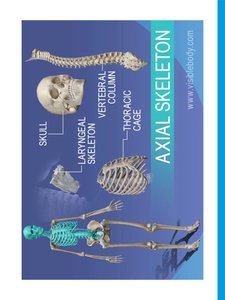
Axial Skeleton
Main Components
The axial skeleton consists of the following structures:
Skull
Laryngeal skeleton
Vertebral column
Thoracic cage
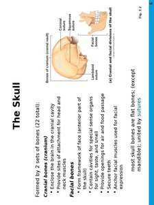
The Skull
Structure and Function
The skull is formed by two sets of bones (22 in total):
Cranial bones (cranium): Enclose the brain in the cranial cavity and provide sites of attachment for head and neck muscles.
Facial bones: Form the framework of the face, contain cavities for special sense organs, provide openings for air and food passage, secure teeth, and anchor facial muscles for expression.
Most skull bones are flat bones (except the mandible) and are united by sutures.
Cranium: Structure
The cranium is divided into two parts:
Cranial vault (calvaria): Forms the superior, lateral, and posterior aspects of the skull.
Cranial base: Forms the skull's floor, containing three fossae (anterior, middle, posterior) that house the brain.
The cranium also contains several named openings for the passage of nerves and blood vessels.
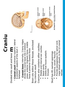
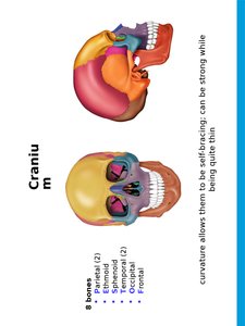
Cranial Bones
There are eight cranial bones:
Parietal (2)
Temporal (2)
Frontal (1)
Occipital (1)
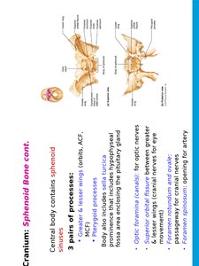
Sphenoid (1)
Ethmoid (1)
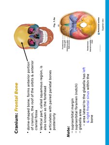
Frontal Bone
Dome-shaped bone forming the anterior portion of the cranium and the roof of the orbits.
Contains the frontal sinuses and forms part of the cranial fossa.
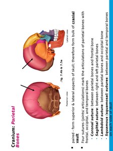
Parietal Bones
Paired bones forming the superior and lateral aspects of the skull.
Articulate with other cranial bones at four main sutures: coronal, sagittal, lambdoid, and squamous.
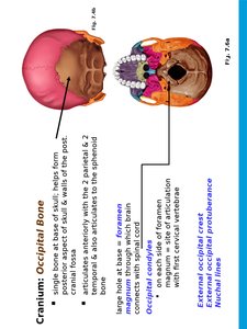
Occipital Bone
Single bone at the base of the skull, forming the posterior cranial fossa.
Contains the foramen magnum, occipital condyles, and external occipital protuberance.
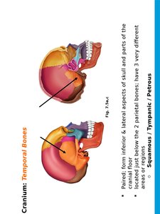
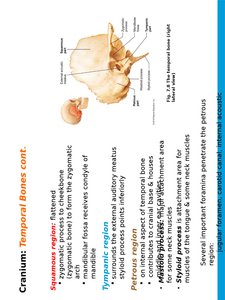
Temporal Bones
Paired bones forming the inferior and lateral aspects of the skull.
Divided into squamous, tympanic, and petrous regions.
Sphenoid Bone
Complex, bat-shaped bone forming the base of the middle cranial fossa and contributing to the base of the skull.
Considered the keystone bone because it articulates with all other cranial bones.
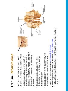
Ethmoid Bone
Deepest skull bone, forming part of the nasal septum and the roof of the nasal cavity.
Contains cribriform plates, perpendicular plate, and ethmoidal air cells.
Major Cranial Sutures
Sutures are immovable joints that connect cranial bones. The four main sutures are:
Coronal: Frontal and parietal bones
Sagittal: Between parietal bones
Lambdoid: Parietal and occipital bones
Squamous: Parietal and temporal bones
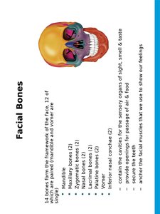
Facial Bones
The facial skeleton consists of 14 bones, including the mandible, maxillae, zygomatic, nasal, lacrimal, palatine, vomer, and inferior nasal conchae. These bones form the framework of the face and provide cavities for the sense organs.

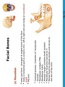
Mandible
Strongest and largest bone of the face.
Forms the lower jaw and contains the lower teeth.
Articulates with the temporal bone at the temporomandibular joint (TMJ).
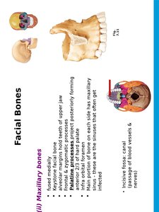
Maxillary Bones
Form the upper jaw and central part of the facial skeleton.
Contain maxillary sinuses and form part of the hard palate.
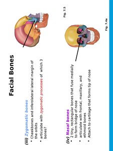
Zygomatic and Nasal Bones
Zygomatic bones: Form the cheekbones and part of the lateral wall of the orbits.
Nasal bones: Form the bridge of the nose.
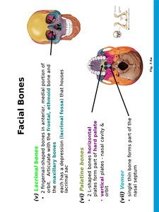
Lacrimal, Palatine, Vomer, and Inferior Nasal Conchae
Lacrimal bones: Form part of the medial wall of the orbits.
Palatine bones: Form part of the hard palate and nasal cavity.
Vomer: Forms part of the nasal septum.
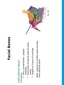
Inferior nasal conchae: Form part of the lateral walls of the nasal cavity.
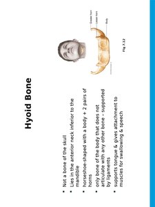
Hyoid Bone
The hyoid bone is a unique, horseshoe-shaped bone in the anterior neck. It does not articulate directly with any other bone and serves as an attachment site for tongue and neck muscles involved in swallowing and speech.
Orbits and Nasal Cavity
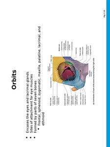
Orbits: Bony cavities that encase the eyes and provide attachment for eye muscles. Formed by parts of seven bones.
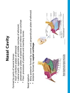
Nasal cavity: Formed by several bones and divided by the nasal septum. Contains the nasal conchae, which increase surface area and help warm and humidify air.
Paranasal Sinuses
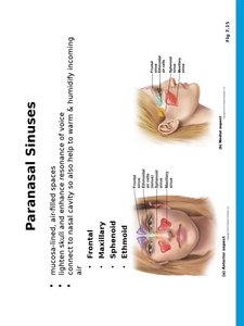
Paranasal sinuses are mucosa-lined, air-filled spaces in the frontal, maxillary, sphenoid, and ethmoid bones. They lighten the skull, enhance voice resonance, and help warm and humidify incoming air.
The Vertebral Column
Structure and Function
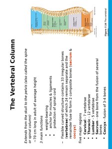
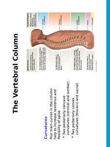
The vertebral column, or spine, extends from the skull to the pelvis and consists of 26 irregular bones. It provides support, protects the spinal cord, and serves as an attachment point for ribs and muscles.
Main regions: Cervical (7), thoracic (12), lumbar (5), sacrum (1, fused), coccyx (1, fused).
Main functions: Transmits body weight, protects the spinal cord, and provides attachment for muscles and ligaments.
Curvatures of the Vertebral Column
The vertebral column has four main curvatures that increase resilience and flexibility:
Cervical and lumbar: Posteriorly concave
Thoracic and sacral: Posteriorly convex
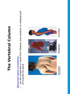
Abnormal Spinal Curvatures
Abnormal curvatures can result from congenital defects, disease, poor posture, or unequal muscle pull. Common types include:
Scoliosis: Lateral curvature
Kyphosis: Exaggerated thoracic curvature
Lordosis: Exaggerated lumbar curvature
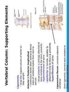
Supporting Elements of the Vertebral Column
Ligaments: Support the column and prevent hyperextension or hyperflexion.
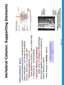
Intervertebral discs: Cushion-like pads between vertebrae, composed of a nucleus pulposus and anulus fibrosus.
Typical Vertebral Structure
All vertebrae share a common structural pattern:
Body (centrum): Weight-bearing region
Vertebral arch: Composed of pedicles and laminae
Vertebral foramen: Canal for the spinal cord
Processes: Spinous, transverse, and articular processes for muscle and ligament attachment
Summary Table: Major Bones of the Skull
Bone | Location | Key Features |
|---|---|---|
Frontal | Forehead, roof of orbits | Frontal sinuses, supraorbital margin |
Parietal (2) | Superior/lateral skull | Articulate at sagittal suture |
Occipital | Posterior skull/base | Foramen magnum, occipital condyles |
Temporal (2) | Inferior/lateral skull | Mastoid/styloid processes, external acoustic meatus |
Sphenoid | Base of skull | Sella turcica, keystone bone |
Ethmoid | Medial orbits, nasal cavity | Cribriform plate, perpendicular plate |
