 Back
BackThe Human Skeleton: Structure, Tissues, and Functions
Study Guide - Smart Notes
 Tailored notes based on your materials, expanded with key definitions, examples, and context.
The Human Skeleton
Introduction
The human skeleton is a complex framework of bones and connective tissues that provides support, protection, and movement for the body. It is composed of various types of tissues, each with specialized functions essential for maintaining homeostasis and facilitating bodily functions.
Functions of the Skeleton
Support: Provides a rigid framework that supports the soft tissues of the body.
Mineral Reservoir: Stores essential minerals such as calcium and phosphate, which can be released into the bloodstream as needed.
Energy Storage: Yellow bone marrow stores lipids, serving as an energy reserve.
Blood Cell Production: Red bone marrow is the site of hematopoiesis, the production of blood cells.
Protection: Shields vital organs (e.g., skull protects the brain, rib cage protects the heart and lungs).
Leverage & Movement: Acts as levers for muscles to produce movement.
Connective Tissues of the Skeleton
Main Types
Connective Tissue Proper: Includes ligaments, bone sheaths, and tendons, which connect bones and muscles.
Supporting Connective Tissue: Includes cartilage (joint discs, intervertebral discs, symphysis pubis) and osseous tissue (bone).
Fluid Connective Tissue: Blood, formed in red bone marrow, circulates nutrients and cells throughout the body.
Developmental Origin: Mesenchyme
All connective tissues of the skeleton originate from mesenchyme, an embryonic connective tissue. Mesenchymal cells differentiate into various cell types:
Fibroblasts/Fibrocytes: Form connective tissue proper.
Adipocytes: Store fat.
Chondroblasts/Chondrocytes: Form cartilage.
Osteoblasts/Osteocytes: Form bone.
Cartilage
Characteristics
Avascular and Aneural: Lacks blood vessels and nerves; nutrients diffuse from surrounding tissues.
High Water Content: Composed of 60–80% water, providing resilience.
Surrounded by Perichondrium: A dense connective tissue layer (except in fibrocartilage) that aids in growth and repair.
Growth and Healing: Cartilage growth ends around 18–20 years; limited healing capacity in adulthood.
Collagen Fibers: Provide tensile strength but poor resistance to shearing forces.
Cartilage Cells
Chondroblasts: Produce the cartilage matrix.
Chondrocytes: Mature cartilage cells located in lacunae within the matrix.
Types of Cartilage
Type | Main Features | Location |
|---|---|---|
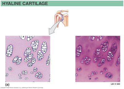
Hyaline Cartilage | Most abundant; fine collagen fibers (not visible); provides support with flexibility | Articular surfaces, costal cartilages, nose, trachea, larynx |
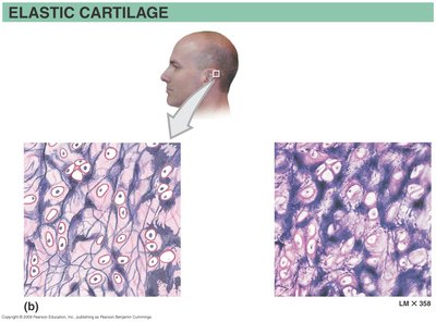
Elastic Cartilage | Many elastic fibers; tolerates repeated bending and recoil | External ear, epiglottis |
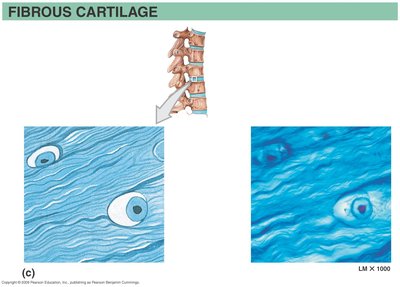
Fibrocartilage | Abundant collagen fibers; resists compression and twisting; lacks perichondrium | Intervertebral discs, pubic symphysis, menisci of knee |

Osseous Tissue (Bone)
Gross Anatomy
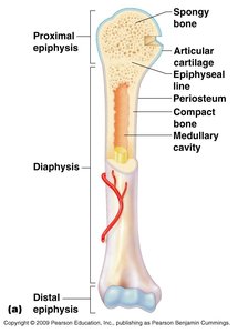
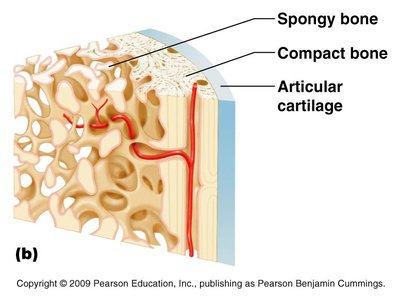
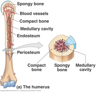
Each bone contains blood vessels, nerves, and lymphatic vessels, and is composed of two main types of osseous tissue: spongy bone and compact bone.

Types of Osseous Tissue
Spongy (Cancellous) Bone:
Honeycomb-like network forming trabeculae (beams) to resist directional stresses.
Cavities filled with red bone marrow; found in the epiphyses and center of bone organs.
Surrounded by endosteum.
Transmits weight and allows for compression and tension along the diaphysis.
Compact Bone:
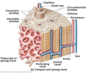
Forms concentric rings of matrix called osteons.
Bone cells are trapped within lacunae.
Organized in nested rings at the periphery of the bone.
Provides strength for weight bearing and protection.



Bone Adaptability
Depending on physiological signals, bone can remodel and change from spongy bone to compact bone and vice versa, allowing adaptation to mechanical stress and metabolic needs.
Summary Table: Cartilage and Bone Comparison
Feature | Cartilage | Bone |
|---|---|---|
Cells | Chondrocytes in lacunae | Osteocytes in lacunae |
Matrix | Gel-like, high water content | Rigid, calcified |
Blood Supply | Avascular | Vascular |
Growth | Appositional and interstitial (until adulthood) | Appositional only |
Healing | Poor | Good |
Key Terms and Definitions
Trabeculae: The lattice-like beams in spongy bone that provide structural support.
Osteon: The structural unit of compact bone, consisting of concentric lamellae around a central canal.
Lacunae: Small spaces within the bone or cartilage matrix that house cells (osteocytes or chondrocytes).
Perichondrium: Dense connective tissue surrounding cartilage (except fibrocartilage).
Endosteum: Thin vascular membrane lining the inner surface of bone.
Periosteum: Dense connective tissue covering the outer surface of bone.
Relevant Equations
While bone structure is not typically described by equations, the following formula is relevant for understanding bone strength:
Stress (σ) = Force (F) / Area (A)
This equation helps explain how bones resist mechanical loads.
