 Back
BackThe Immune System: Complement Cascade, Blood Groups, and Hypersensitivity
Study Guide - Smart Notes
 Tailored notes based on your materials, expanded with key definitions, examples, and context.
The Immune System
The Complement Cascade
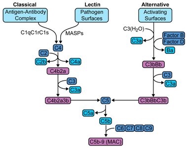
The complement system is a crucial part of innate and adaptive immunity, consisting of a series of plasma proteins that, when activated, enhance the ability of antibodies and phagocytic cells to clear microbes and damaged cells. It can be activated via three main pathways: classical, lectin, and alternative.
Classical Pathway: Triggered by antigen-antibody complexes, involving C1q, C1r, and C1s proteins.
Lectin Pathway: Initiated by mannose-binding lectin binding to pathogen surfaces, activating MASPs.
Alternative Pathway: Activated directly by pathogen surfaces, involving factors B and D.
All pathways converge at the activation of C3, leading to the formation of the membrane attack complex (MAC).
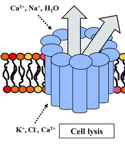
Key Function: The MAC forms pores in the target cell membrane, causing cell lysis by osmolysis.
Membrane Attack Complex (MAC)
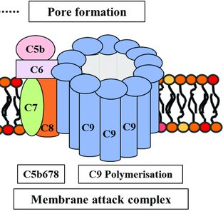
The MAC is the cytolytic effector of the complement system. It forms giant pores in the plasma membrane of pathogens or targeted cells, leading to their destruction.
Formation: Sequential assembly of complement proteins C5b, C6, C7, C8, and multiple C9 molecules.
Mechanism: The MAC inserts into the cell membrane, creating a pore that disrupts cellular homeostasis.
Result: Influx of water and ions (Ca2+, Na+, H2O) and efflux of K+, Cl-, leading to cell swelling and lysis.
Blood Transfusions and Blood Groups
ABO and Rh Blood Groups
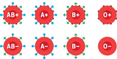
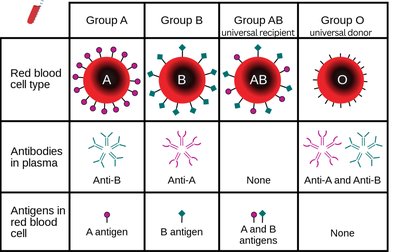
Red blood cells (RBCs) express specific antigens on their surface that determine blood type. The two main systems are ABO and Rh.
ABO System: Four main blood types: A, B, AB, and O, based on the presence or absence of A and B antigens.
Rh System: Presence (+) or absence (−) of the RhD antigen.
Genetics: ABO alleles are inherited from parents; A and B are dominant over O. RhD+ is dominant over RhD−.
Blood Transfusion Compatibility
Transfusion reactions occur if incompatible blood is transfused, leading to antibody-mediated RBC destruction. Compatibility depends on both ABO and Rh antigens.
Antibodies: Individuals produce antibodies against ABO antigens they do not possess.
Universal Donor: Type O− (no A, B, or Rh antigens).
Universal Recipient: Type AB+ (all antigens present, no anti-A, anti-B, or anti-Rh antibodies).
Recipient | A donor | B donor | AB donor | O donor |
|---|---|---|---|---|
A | ✓ | ✗ | ✓ | ✗ |
B | ✗ | ✓ | ✓ | ✗ |
AB | ✓ | ✓ | ✓ | ✓ |
O | ✗ | ✗ | ✗ | ✓ |
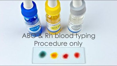
Antibody Typing Test
Blood typing is performed in the laboratory using specific antibodies to detect the presence of A, B, and Rh antigens on RBCs. Agglutination indicates a positive reaction.
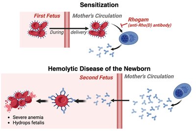
Hemolytic Disease of the Newborn (HDN)
HDN occurs when an RhD− mother carries an RhD+ fetus. Sensitization during the first pregnancy leads to maternal anti-RhD antibody production, which can attack fetal RBCs in subsequent pregnancies, causing anemia and other complications.
Prevention: Administration of RhoGAM (anti-RhD antibody) prevents maternal sensitization.
Immune Response to Allergens (Hypersensitivity)
Allergy and Hypersensitivity
An allergy is an immune response to a non-pathogenic exogenous antigen (allergen), such as pollen, food, or animal dander. Hypersensitivity reactions can be immediate or delayed.
Sensitization Phase: First exposure to allergen leads to immune priming.
Immediate Hypersensitivity: Occurs within minutes, mediated by IgE antibodies and mast cells (e.g., anaphylaxis).
Delayed Hypersensitivity: Occurs within days, mediated by T helper cells and macrophages.
Mast Cells: Tissue basophils that release histamine and other mediators upon activation.
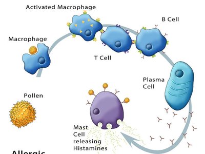
Mechanism of Allergic Response
Upon re-exposure to the allergen, memory B cells produce IgE, which binds to mast cells. Allergen cross-linking triggers mast cell degranulation, releasing histamine and other mediators, causing vasodilation, bronchoconstriction, and inflammation.
Key Steps:
Allergen presentation by APCs on MHC-II
Activation of T helper and B cells
IgE production and binding to mast cells
Mast cell degranulation and mediator release
Clinical Manifestations: Local or systemic allergic reactions, including anaphylaxis.
Summary Tables
Red blood cell type | Group A | Group B | Group AB | Group O |
|---|---|---|---|---|
Antibodies in plasma | Anti-B | Anti-A | None | Anti-A and Anti-B |
Antigens in red blood cell | A antigen | B antigen | A and B antigens | None |
Additional info: The complement system also plays a role in inflammation and opsonization, enhancing phagocytosis of pathogens. The Rh system is clinically significant in pregnancy and transfusion medicine due to the risk of hemolytic disease of the newborn.
