 Back
BackThe Immune System: Innate and Adaptive Defenses
Study Guide - Smart Notes
 Tailored notes based on your materials, expanded with key definitions, examples, and context.
Introduction to the Immune System and Infectious Agents
Overview of the Immune System
The immune system is a complex network of cells and molecules distributed throughout the body, unified by their function to provide immunity—protection from harmful agents. Unlike other body systems, it is not defined by specific organs but by its cellular and molecular components.
Immunity: The ability to resist or eliminate potentially harmful foreign materials or abnormal cells.
Infectious agents (pathogens): Microorganisms that can cause disease, including bacteria, viruses, fungi, protozoans, and multicellular parasites.
Categories of Infectious Agents
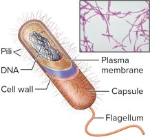
Bacteria: Single-celled prokaryotes lacking a nuclear envelope, enclosed by a cell wall. They can cause disease by releasing toxins or invading tissues.
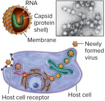
Viruses: Non-cellular infectious agents consisting of DNA or RNA within a protein shell (capsid). They are obligate intracellular parasites, requiring host cells to reproduce.
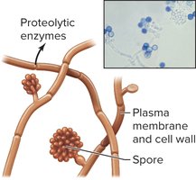
Fungi: Eukaryotic organisms with a plasma membrane and cell wall. Includes molds, yeasts, and multicellular fungi that produce spores. They release proteolytic enzymes that induce inflammation.
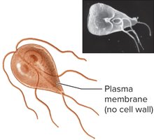
Protozoans: Eukaryotic cells without a cell wall. Disease-causing protozoans are called parasites (e.g., malaria, trichomoniasis).
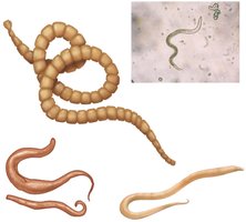
Multicellular Parasites: Nonmicroscopic organisms such as parasitic worms (e.g., tapeworms) that derive nourishment from their host.
Prions: Infectious protein fragments that cause diseases in nervous tissue (e.g., Variant Creutzfeldt-Jakob disease).
Overview of the Immune System
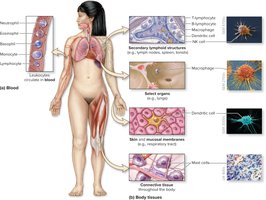
Immune Cells and Their Locations
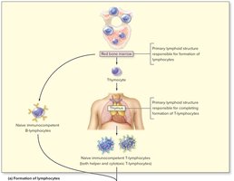
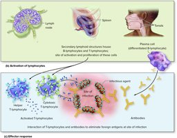
Immune cells, primarily leukocytes (white blood cells), are formed in the red bone marrow and distributed throughout the body. They are housed in various tissues and organs, including secondary lymphoid structures (lymph nodes, spleen, tonsils, MALT), select organs, skin, mucosal membranes, and connective tissue.
Granulocytes: Neutrophils, eosinophils, basophils
Monocytes: Become macrophages in tissues
Lymphocytes: B-lymphocytes, T-lymphocytes, NK cells
Cytokines
Cytokines are small proteins that regulate immune activity. They act as chemical messengers, influencing the behavior and development of immune cells, regulating inflammation, and signaling between cells. Cytokines can act in autocrine, paracrine, or endocrine manners and have a short biological half-life.
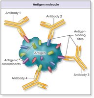
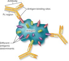
Antigens and Antigenic Determinants
An antigen is a unique molecule (often a protein or polysaccharide) capable of binding to components of the adaptive immune system. Lymphocytes recognize specific regions of antigens called antigenic determinants or epitopes. Immunogens are antigens that induce an immune response. Haptens are small molecules that become immunogenic only when attached to a larger carrier molecule.
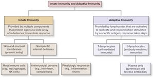
Comparison of Innate and Adaptive Immunity
The immune system is divided into two main branches:
Innate Immunity: Nonspecific, present at birth, provides immediate defense against a wide range of pathogens. Includes physical barriers (skin, mucous membranes), cellular defenses, and molecular defenses (e.g., interferons, complement).
Adaptive Immunity: Specific, acquired through exposure to antigens, involves T- and B-lymphocytes. Provides a targeted response and immunologic memory.
Innate Immunity
First Line of Defense: Physical and Chemical Barriers
Skin: Keratinized stratified squamous epithelium and antimicrobial secretions (e.g., dermicidin, lysozyme, sebum, defensins) prevent pathogen entry.
Mucous Membranes: Line body openings, produce mucus, and release antimicrobial substances. Cilia, saliva, stomach acid, and acidic secretions further protect against pathogens.
Microbiome: Nonpathogenic microorganisms that reside on body surfaces and compete with pathogens.
Second Line of Defense: Nonspecific Internal Defenses
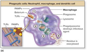
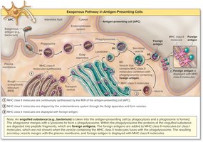
Phagocytic Cells: Neutrophils, macrophages, and dendritic cells engulf and destroy pathogens. Dendritic cells also present antigens to T-lymphocytes, initiating adaptive immunity.
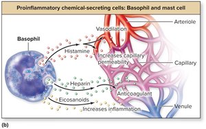
Proinflammatory Chemical-Secreting Cells: Basophils and mast cells release histamine, heparin, and eicosanoids to promote inflammation and attract immune cells.
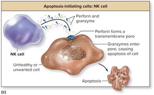
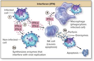
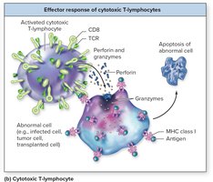
Apoptosis-Initiating Cells: NK (natural killer) cells induce apoptosis in virus-infected, tumor, or abnormal cells by releasing perforin and granzymes.
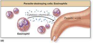
Eosinophils: Attack multicellular parasites by releasing cytotoxic chemicals and participate in allergic responses.
Antimicrobial Proteins
Interferons (IFNs): Cytokines that interfere with viral replication and activate NK cells and macrophages.
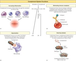
Complement System: A group of plasma proteins that enhance immune responses through inflammation, opsonization, cytolysis, and elimination of immune complexes.
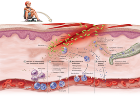
Inflammation
Inflammation is an immediate, local, nonspecific response to injury or infection. It involves the release of inflammatory chemicals, vascular changes (vasodilation, increased permeability), recruitment of leukocytes, and delivery of plasma proteins. The cardinal signs are redness, heat, swelling, pain, and loss of function.
Fever
Fever (pyrexia) is an elevation of body temperature due to pyrogens. It inhibits microbial reproduction, enhances immune responses, and accelerates tissue repair. However, high fevers can be dangerous and cause protein denaturation or brain damage.
Adaptive Immunity
Branches of Adaptive Immunity
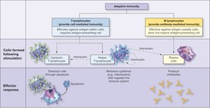
Adaptive immunity is the third line of defense and involves specific lymphocyte responses to antigens. It is divided into:
Cell-mediated immunity: Involves T-lymphocytes (helper and cytotoxic T-cells).
Antibody-mediated (humoral) immunity: Involves B-lymphocytes, plasma cells, and antibodies.
Structure and Function of Lymphocytes
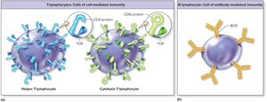
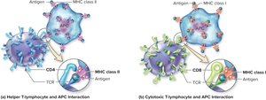
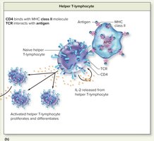
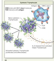
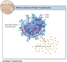
T-lymphocytes: Have T-cell receptors (TCRs) and CD molecules (CD4 on helper T-cells, CD8 on cytotoxic T-cells). Helper T-cells assist other immune cells; cytotoxic T-cells destroy infected or abnormal cells.
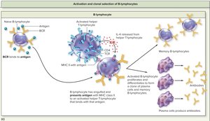
B-lymphocytes: Have B-cell receptors (BCRs) and directly bind antigens. Differentiate into plasma cells (antibody producers) or memory B-cells.
Antigen Presentation and MHC Molecules
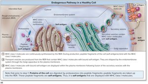
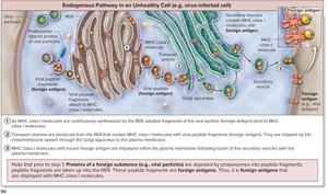
MHC I: Found on all nucleated cells; presents endogenous antigens to cytotoxic T-cells.
MHC II: Found on antigen-presenting cells (APCs); presents exogenous antigens to helper T-cells.
Lymphocyte Life Events
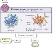
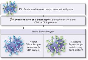
Formation and Selection: Occurs in primary lymphoid organs (red marrow, thymus). Lymphocytes become immunocompetent and self-tolerant.
Activation: Occurs in secondary lymphoid structures upon antigen exposure, leading to clonal selection and proliferation.
Effector Response: Lymphocytes act to eliminate the antigen (T-cells migrate to infection site; B-cells produce antibodies).
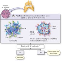
Selection and Differentiation of T-lymphocytes
Positive Selection: T-cells that can bind MHC molecules survive.
Negative Selection: T-cells that bind self-antigens are eliminated (central tolerance).
Differentiation: Helper T-cells retain CD4; cytotoxic T-cells retain CD8.
Activation and Effector Response of Lymphocytes
Activation of Helper and Cytotoxic T-Lymphocytes
Helper T-cells: Activated by antigen presentation with MHC II on APCs; secrete IL-2 to stimulate themselves and other immune cells.
Cytotoxic T-cells: Activated by antigen presentation with MHC I on APCs; require IL-2 from helper T-cells for full activation.
Activation of B-Lymphocytes
B-cells bind intact antigen, process and present it to helper T-cells, and are stimulated by IL-4 to proliferate and differentiate into plasma cells and memory B-cells.
Effector Responses
Helper T-cells: Release cytokines to regulate both innate and adaptive immune responses.
Cytotoxic T-cells: Induce apoptosis in infected or abnormal cells by releasing perforin and granzymes.
Plasma cells: Produce and secrete antibodies that circulate in blood and lymph.
Antibodies (Immunoglobulins)
Structure of Immunoglobulins
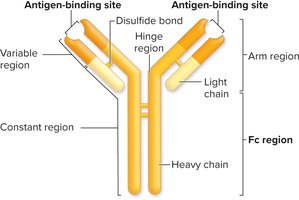
Antibodies are Y-shaped proteins composed of two heavy and two light chains, with variable regions forming antigen-binding sites and a constant (Fc) region determining biological function.
Actions of Antibodies
Neutralization: Antibody covers antigenic determinant, preventing pathogen activity.
Agglutination: Antibody cross-links antigens, causing clumping.
Precipitation: Antibody cross-links soluble antigens, forming insoluble complexes.
Complement Activation: Fc region binds complement proteins, triggering classical pathway.
Opsonization: Fc region enhances phagocytosis by binding to phagocyte receptors.
Activation of NK Cells: Fc region triggers NK cell-mediated cytotoxicity.
Classes of Immunoglobulins
IgG: Most abundant, crosses placenta, participates in all antibody actions.
IgM: First produced, effective at agglutination and complement activation.
IgA: Found in secretions, protects mucosal surfaces.
IgD: Functions as B-cell receptor.
IgE: Involved in allergic reactions and defense against parasites.
Immunologic Memory and Immunity Types
Immunologic Memory
Memory cells formed during the primary immune response enable a faster and stronger secondary response upon re-exposure to the same antigen. This principle underlies the effectiveness of vaccines.
Active and Passive Immunity
Active Immunity: Results from direct exposure to antigen (infection or vaccination); produces memory cells and long-term protection.
Passive Immunity: Results from transfer of antibodies (e.g., maternal antibodies, antiserum); does not produce memory cells or long-term protection.
