 Back
The Immune System: Innate and Adaptive Defenses
Study Guide - Smart Notes
 Tailored notes based on your materials, expanded with key definitions, examples, and context.
The Immune System: Innate and Adaptive Defenses
Overview of Immune System Function
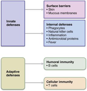
The immune system provides resistance to disease through a complex network of cells, proteins, and mechanisms. It is divided into two main branches: innate (nonspecific) defenses and adaptive (specific) defenses. These systems work together to protect the body from pathogens and other harmful agents.
Innate (Nonspecific) Defenses
First Line of Defense: Surface Barriers
The first line of defense consists of physical and chemical barriers that prevent the entry of pathogens into the body. These include the skin, mucous membranes, and their secretions.
Skin: Acts as a physical barrier; keratin in the skin is resistant to weak acids, bases, and bacterial enzymes.
Mucous membranes: Line body cavities open to the exterior and trap microorganisms.
Protective chemicals: Include acid mantle, enzymes (e.g., lysozyme), mucin, defensins, and other antimicrobial substances.
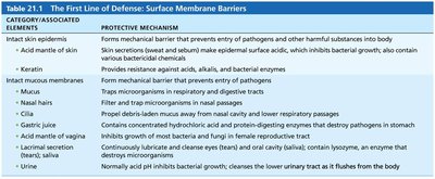
Respiratory system modifications include mucus-coated hairs in the nose and cilia in the upper respiratory tract, which help trap and remove pathogens.
Second Line of Defense: Internal Defenses
If pathogens breach surface barriers, the innate system employs internal defenses such as phagocytes, natural killer cells, inflammation, antimicrobial proteins, and fever.
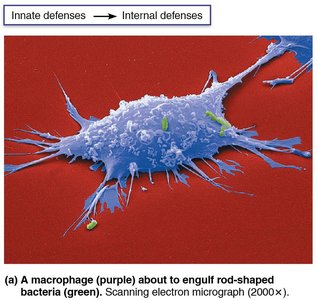
Phagocytes: White blood cells that ingest and digest foreign invaders. Main types include neutrophils and macrophages.
Natural Killer (NK) cells: Lymphocytes that target and kill virus-infected and cancerous cells by inducing apoptosis.
Inflammation: A tissue response to injury that prevents the spread of damaging agents, disposes of pathogens, and sets the stage for repair.
Antimicrobial proteins: Interferons and complement proteins that attack microorganisms or hinder their reproduction.
Fever: Systemic response that inhibits microbial growth and speeds up tissue repair.
Phagocytosis: Steps and Mechanisms
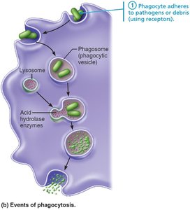
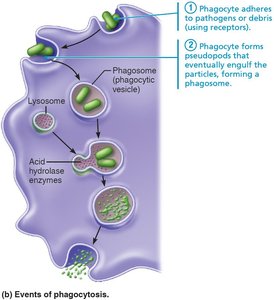
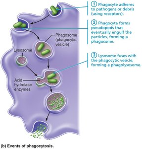
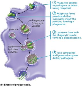
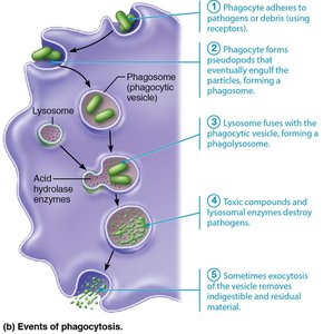
Phagocytosis is the process by which phagocytes engulf and destroy pathogens. The steps include:
Recognition and adherence to the pathogen's surface.
Engulfment of the pathogen into a phagosome.
Fusion of the phagosome with a lysosome to form a phagolysosome.
Destruction of the pathogen by lysosomal enzymes and toxic compounds.
Exocytosis of indigestible material.
Inflammation: Tissue Response to Injury
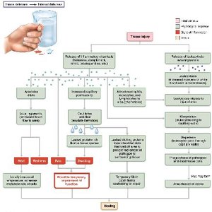
Inflammation is triggered by tissue injury and serves to contain damage, remove pathogens, and initiate repair. The four cardinal signs are redness, heat, swelling, and pain. The process involves:
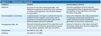
Release of inflammatory chemicals (e.g., histamine, kinins, prostaglandins, complement, cytokines).
Vasodilation and increased vascular permeability, leading to edema.
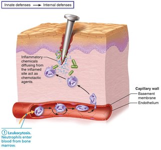
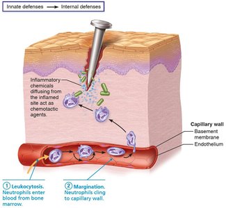
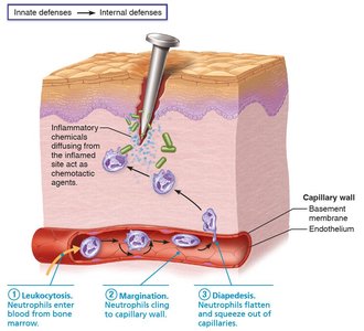
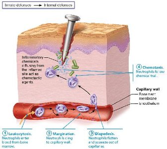
Phagocyte mobilization to the site of injury.
Phagocyte Mobilization Steps
Leukocytosis: Neutrophils enter blood from bone marrow.
Margination: Neutrophils cling to capillary walls.
Diapedesis: Neutrophils squeeze out of capillaries into tissues.
Chemotaxis: Neutrophils follow chemical signals to the site of injury.
Antimicrobial Proteins
Interferons
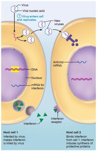
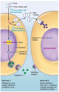
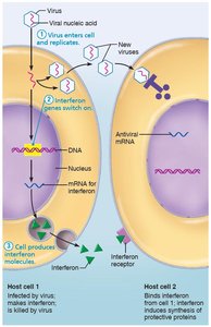
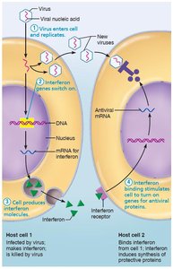
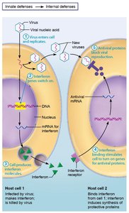
Interferons (IFNs) are proteins released by virus-infected cells that help protect neighboring cells from viral infection. They stimulate the production of antiviral proteins and activate immune cells such as NK cells and macrophages.
Complement System
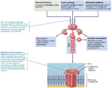
The complement system is a group of about 20 plasma proteins that enhance the ability of antibodies and phagocytic cells to clear microbes and damaged cells. It can be activated by three pathways:
Classical pathway: Triggered by antibodies binding to antigens.
Lectin pathway: Initiated by lectins binding to microbial carbohydrates.
Alternative pathway: Activated spontaneously on microbial surfaces.
All pathways converge on the activation of C3, leading to opsonization, inflammation, and cell lysis via the membrane attack complex (MAC).
Fever
Fever is a systemic response to infection, mediated by pyrogens released from leukocytes and macrophages. It increases body temperature, which inhibits microbial growth and accelerates tissue repair.
Summary Table: Second Line of Defense
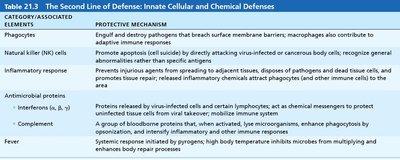
Additional info: The innate and adaptive immune systems are highly integrated, with many molecules and cells participating in both arms of defense. For example, phagocytes not only destroy pathogens but also present antigens to adaptive immune cells, linking the two systems.
