 Back
The Immune System: Innate and Adaptive Body Defenses
Study Guide - Smart Notes
 Tailored notes based on your materials, expanded with key definitions, examples, and context.
The Immune System: Innate and Adaptive Body Defenses
Introduction to the Immune System
The immune system is a functional system that protects the body from disease by distinguishing self from non-self and eliminating pathogens and abnormal cells. It is essential for maintaining homeostasis and preventing disease.
Self vs. Non-self Recognition: The immune system can identify and target foreign invaders such as bacteria, viruses, fungi, and cancer cells.
Functional System: Unlike organ systems, the immune system is composed of cells and molecules distributed throughout the body.
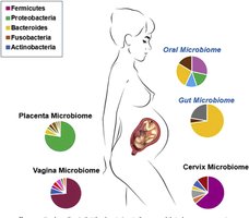
Innate (Nonspecific) Defenses
Overview of Innate Immunity
Innate immunity provides the first and second lines of defense against pathogens. It is present from birth and responds rapidly to a wide range of invaders.
First Line: External body membranes (skin and mucosae)
Second Line: Internal defenses including phagocytes, natural killer cells, antimicrobial proteins, inflammation, and fever
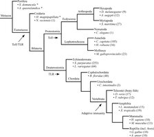
Evolutionary Context of Innate Immunity
Innate immunity is an ancient defense mechanism, present across many animal phyla, and is germline-encoded.
Evolution: Genes for innate immunity are conserved from insects to vertebrates, originating over 600 million years ago.
First Line of Defense: Surface Barriers
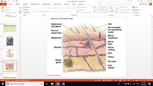
The skin and mucous membranes act as physical and chemical barriers to infection.
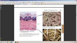
Epidermis: Multiple layers of keratinized cells resist penetration and shedding removes microbes.
Dermis: Collagen fibers provide mechanical strength.
Chemical Defenses: Sweat (acidic pH, salt, dermcidin), sebum (lowers pH), lysozyme in saliva and tears, and mucus trap and destroy microbes.
Other First-Line Defenses
Antimicrobial Peptides: Destroy microbes directly.
Secretions: Saliva, stomach acid, bile, urine, and vaginal secretions wash away or destroy pathogens.
Physical Processes: Peristalsis, defecation, vomiting, menstrual flow, and blood flow help expel microbes.
Second Line of Defense: Internal Defenses
When pathogens bypass surface barriers, internal defenses are activated.
Phagocytes: Macrophages and neutrophils ingest and destroy pathogens.
Natural Killer (NK) Cells: Destroy infected or abnormal cells by inducing apoptosis.
Antimicrobial Proteins: Interferons and complement proteins inhibit pathogen spread.
Inflammation: Local tissue response to injury or infection.
Fever: Systemic response that inhibits pathogen growth and speeds repair.
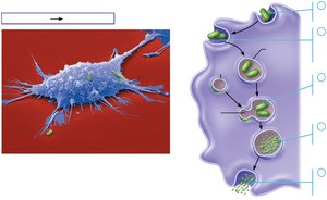
Phagocytosis
Phagocytes engulf and digest pathogens through a multi-step process.
Steps: Adhesion, ingestion, maturation (phagolysosome formation), killing, and elimination.
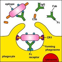
Opsonization: Coating of pathogens with antibodies or complement to enhance phagocytosis.
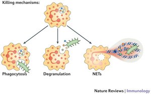
Nonphagocytic Killing (Neutrophils)
Neutrophils can kill microbes by releasing toxic chemicals or forming neutrophil extracellular traps (NETs).
Mechanisms: Phagocytosis, degranulation, and NETosis.
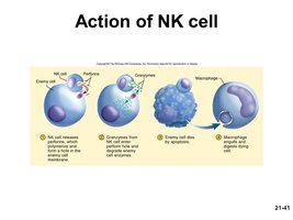
Natural Killer (NK) Cells
NK cells target and destroy virus-infected and cancerous cells by recognizing abnormal surface markers.
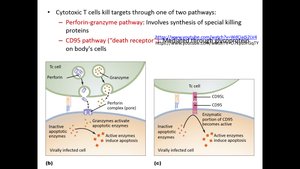
Mechanism: Release of perforins and granzymes to induce apoptosis.
Role: Surveillance in blood and lymph, promoting inflammation.
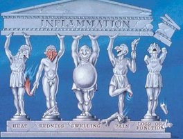
Inflammation
Inflammation is a protective response to tissue injury or infection, characterized by redness, heat, swelling, pain, and loss of function.
Functions: Prevents spread of damage, disposes of debris, and initiates repair.
Cardinal Signs: Redness, heat, swelling, pain, impairment of function.
Vasodilation and Increased Vascular Permeability
Chemical mediators (cytokines, histamine, kinins, prostaglandins, leukotrienes, complement) trigger vasodilation and increased permeability, leading to local hyperemia and edema.
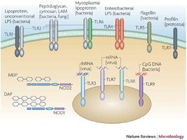
TLRs: Toll-like receptors on macrophages recognize microbial patterns and initiate inflammation.
Phagocyte Mobilization
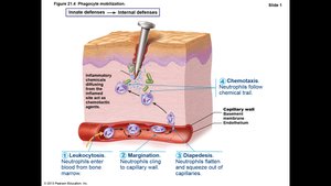
Phagocytes are recruited to sites of inflammation through a series of steps: leukocytosis, margination, diapedesis, and chemotaxis.
Pus Formation: Accumulation of dead cells and pathogens may form pus or abscesses.
Antimicrobial Proteins
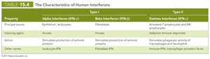
Interferons and complement proteins enhance innate defenses by inhibiting viral replication and promoting pathogen destruction.
Interferons: Released by infected cells to protect neighboring cells.
Complement System: Serum proteins that promote opsonization, chemotaxis, and cell lysis.
Fever
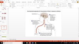
Fever is a systemic response to infection, mediated by pyrogens that reset the hypothalamic thermostat.
Benefits: Inhibits microbial growth, increases metabolic rate, and enhances tissue repair.
Risks: High fever can denature enzymes and be harmful.
Adaptive (Specific) Defenses
Overview of Adaptive Immunity
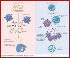
Adaptive immunity is characterized by specificity, memory, systemic action, clonality, and self-tolerance. It is slower to respond but provides long-lasting protection.
Humoral Immunity: Mediated by B cells and antibodies in body fluids.
Cell-mediated Immunity: Mediated by T cells that target infected or abnormal cells.
Antigens
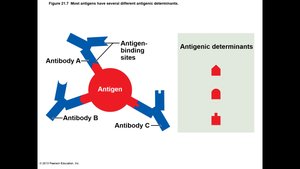
Antigens are substances that trigger adaptive immune responses. They are usually large, complex molecules not normally found in the body.
Types: Exogenous (external), endogenous (internal), and autoantigens (self-derived).
Antigenic Determinants: Specific regions of antigens recognized by antibodies or lymphocyte receptors.
Cells of the Adaptive Immune System
B Lymphocytes (B cells): Responsible for humoral immunity; mature in bone marrow.
T Lymphocytes (T cells): Responsible for cell-mediated immunity; mature in thymus.
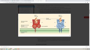
Antigen-Presenting Cells (APCs): Dendritic cells, macrophages, and B cells that present antigens to lymphocytes using MHC molecules.
Humoral Immune Response
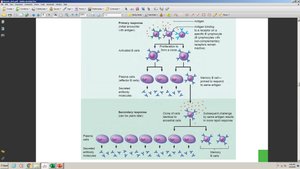
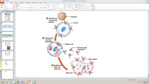
B cells recognize specific antigens via their B cell receptors (BCRs) and, upon activation, differentiate into plasma cells that secrete antibodies.
Primary Response: First exposure to antigen; slower and lower antibody production.
Secondary Response: Subsequent exposures; faster and stronger due to memory cells.
Classes of Antibodies
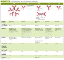
There are five main classes of antibodies (immunoglobulins): IgM, IgG, IgA, IgD, and IgE. Each has distinct roles and properties.
IgM: First antibody produced; effective in agglutination and complement activation.
IgG: Most abundant; crosses placenta; provides long-term immunity.
IgA: Found in mucosal areas and secretions.
IgD: Functions mainly as a B cell receptor.
IgE: Involved in allergic responses and defense against parasites.
Types of Acquired Immunity
Immunity can be acquired actively (through infection or vaccination) or passively (through maternal antibodies or antibody injection).
Type | Active | Passive |
|---|---|---|
Naturally Acquired | Infection | Maternal antibodies |
Artificially Acquired | Vaccination | Antibody injection (e.g., antiserum) |

Mechanisms of Antibody Action
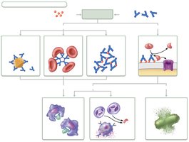
Antibodies neutralize, agglutinate, and precipitate antigens, and activate complement, enhancing phagocytosis and inflammation.
Cell-Mediated Immune Response
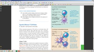
T cells recognize processed antigen fragments presented by MHC molecules on cell surfaces. There are several types of T cells:
Helper T Cells (CD4+): Activate other immune cells via cytokine secretion.
Cytotoxic T Cells (CD8+): Directly kill infected or abnormal cells.
Regulatory T Cells: Suppress immune responses to prevent autoimmunity.
Homeostatic Imbalances of Immunity
Immunodeficiencies
Primary (Congenital): Genetic defects (e.g., SCID) impair immune cell function.
Secondary (Acquired): Result from infections (e.g., AIDS), cancers, or immunosuppressive drugs.
Autoimmune Diseases
Mechanism: Immune system attacks self-antigens, leading to tissue damage (e.g., multiple sclerosis, type 1 diabetes, rheumatoid arthritis).
Treatment: Immunosuppressive drugs and anti-inflammatory agents.
Hypersensitivity Reactions
Hypersensitivities are exaggerated immune responses to harmless antigens (allergens).
Type | Immune Reactant | Antigen | Mechanism | Examples |
|---|---|---|---|---|
I (Immediate) | IgE | Soluble | Mast cell activation | Allergic rhinitis, anaphylaxis |
II (Cytotoxic) | IgG/IgM | Cell-associated | Complement-mediated lysis | Transfusion reaction |
III (Immune Complex) | IgG/IgM | Soluble | Immune complex deposition | Serum sickness, SLE |
IV (Delayed) | T cells | Cell-associated | Cell-mediated cytotoxicity | Contact dermatitis |
Developmental Aspects and Aging of the Immune System
Immune function declines with age due to thymic involution and reduced lymphocyte activity, increasing susceptibility to infections and cancer.
Antibody Changes: IgA and IgG increase, IgM and IgE decrease.
Clinical Implications: Elderly may not tolerate immunosuppressive therapies well.
