 Back
BackThe Immune System: Lines of Defense and Immune Responses
Study Guide - Smart Notes
 Tailored notes based on your materials, expanded with key definitions, examples, and context.
The Immune System
The Three Lines of Defense
The immune system protects the body from pathogens through three main lines of defense: physical barriers, the innate immune response, and the adaptive immune response. Each line of defense involves specific cells, molecules, and mechanisms to prevent and eliminate infections.
Physical Barriers
Types and Functions of Physical Barriers
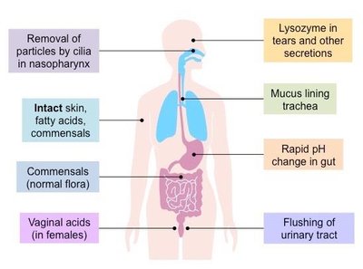
Physical barriers are the body's first line of defense, preventing pathogens from entering tissues and organs. These barriers include the skin, mucous membranes, and various secretions.
Intact skin: Acts as a physical shield; fatty acids and commensal microbes provide additional protection.
Mucus and cilia: Trap and remove particles in the respiratory tract.
Stomach acidity: Destroys ingested pathogens.
Commensal microbiota: Compete with pathogens and promote immune tolerance.
Vaginal acids (in females): Create an acidic environment that inhibits pathogen growth.
Lysozyme in tears and secretions: Enzyme that breaks down bacterial cell walls.
Flushing of urinary tract: Removes microbes from the urinary system.
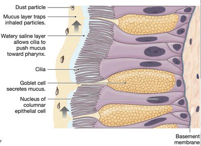
Mucociliary Escalator
The mucociliary escalator is a specialized mechanism in the respiratory tract where mucus traps inhaled particles, and cilia move the mucus toward the pharynx for removal.
Goblet cells: Secrete mucus to trap particles.
Cilia: Beat rhythmically to move mucus and trapped particles out of the airways.
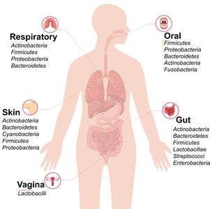
The Human Microbiome
The human microbiome consists of trillions of symbiotic microbes (bacteria, fungi, viruses) living on the skin, gut, respiratory tract, and reproductive organs. These microbes play essential roles in health and immunity.
Colonization: Begins at birth and stabilizes by age three.
Functions: Compete with pathogens, produce antimicrobial agents, aid in nutrient and drug metabolism, and promote immune tolerance.
Disruption: Failure of tolerance can lead to diseases such as inflammatory bowel disease and atopic dermatitis.
Innate Immune Response
Overview and Key Cells
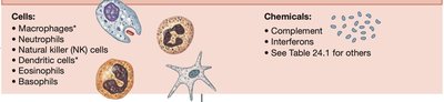
The innate immune response is the body's immediate, non-specific reaction to pathogens. It is not remembered by the immune system and involves various cells and chemicals.
Granulocytes: Neutrophils, eosinophils, basophils
Macrophages and dendritic cells (DCs): Phagocytosis and cytokine secretion
Natural Killer (NK) cells: Kill virus-infected and cancer cells
Chemicals: Complement proteins, interferons, and other cytokines
Key Chemicals of the Innate Immune Response
Various chemicals mediate inflammation, pathogen destruction, and immune cell recruitment.
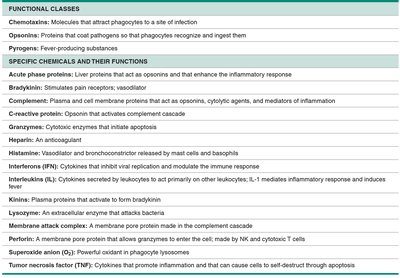
Class | Function |
|---|---|
Chemotaxins | Attract phagocytes to infection sites |
Opsonins | Coat pathogens for easier phagocytosis |
Pyrogens | Induce fever |
Complement | Plasma proteins that mediate inflammation and cell lysis |
Interferons | Inhibit viral replication |
Lysozyme | Enzyme that attacks bacteria |
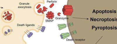
Perforin, Granzymes | Induce apoptosis in target cells |
Phagocytosis
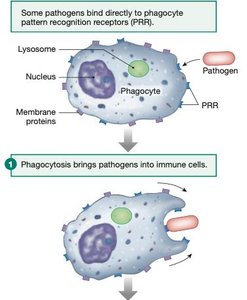
Phagocytosis is the process by which immune cells ingest and destroy pathogens. It is mediated by neutrophils, macrophages, and dendritic cells.
Recognition: Pathogens bind to pattern recognition receptors (PRRs) on phagocytes.
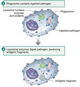
Engulfment: Pathogen is internalized into a phagosome.
Digestion: Lysosomal enzymes digest the pathogen, producing antigenic fragments.
Major Histocompatibility Complex (MHC)
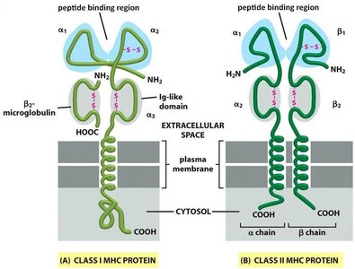
MHC proteins are membrane-bound molecules that present antigen fragments to T cells. There are two main classes:
MHC I: Present on all nucleated cells; present endogenous antigens to cytotoxic T cells.
MHC II: Present on antigen-presenting cells (APCs); present exogenous antigens to helper T cells.
Natural Killer (NK) Cells and Apoptosis
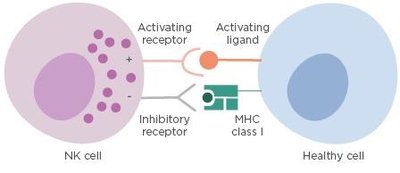
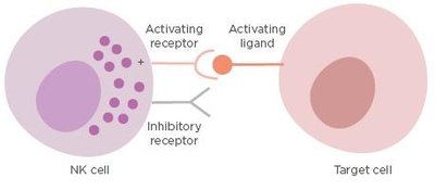
NK cells recognize and kill cells with low MHC I expression, a sign of viral infection or transformation. They induce apoptosis via cytotoxic granules containing perforin and granzymes, or by expressing death ligands.
Healthy cells: Express normal MHC I, inhibiting NK cell activation.
Target cells: Lack MHC I, leading to NK cell-mediated killing.
Complement System
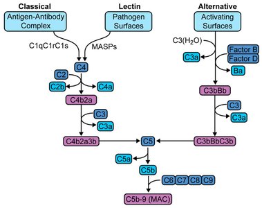
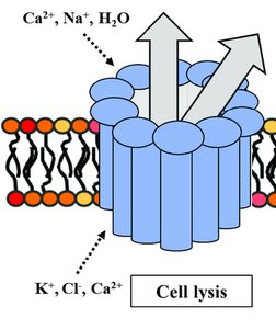
The complement system is a cascade of plasma proteins that enhances the ability of antibodies and phagocytic cells to clear microbes and damaged cells. It can be activated by three pathways: classical, lectin, and alternative.
Membrane Attack Complex (MAC): Forms pores in target cell membranes, causing lysis.
Inflammation
Inflammation is a hallmark of the innate immune response, characterized by redness, heat, swelling, and pain. These signs result from increased blood flow, capillary permeability, and the action of inflammatory mediators.
Redness (rubor): Due to vasodilation.
Heat (calor): Due to increased blood flow.
Swelling (tumor): Due to fluid leakage into tissues.
Pain (dolor): Due to stimulation of nerve endings.
Adaptive Immune Response
Overview and Key Molecules
The adaptive immune response is antigen-specific, generates immune memory, and is mediated by lymphocytes (B and T cells). It is slower to develop but provides long-lasting protection.
B cells: Produce antibodies and mediate humoral immunity.
T cells: Include cytotoxic T cells (kill infected cells), helper T cells (activate other immune cells), and regulatory T cells (suppress immune responses).
Antibodies: Secreted immunoglobulins that bind specific antigens.
T cell receptors (TCRs): Recognize antigen-MHC complexes.
Lymphocyte Activation and Clonal Expansion
Naive lymphocytes are mature B and T cells that have not yet encountered their specific antigen. Upon antigen exposure, they become activated, proliferate, and differentiate into effector and memory cells.
Clonal expansion: Rapid proliferation of antigen-specific lymphocytes.
Memory cells: Provide faster and stronger responses upon re-exposure to the same antigen.
Antibody Structure and Function
Antibodies are Y-shaped molecules composed of two heavy and two light chains. They can be membrane-bound (B cell receptors) or secreted (immunoglobulins).
Antigen-binding sites: Located at the tips of the Y arms; bind specific antigens.
Fc region: Interacts with immune cells and complement proteins.
Functions: Neutralization, opsonization, complement activation, and agglutination.
Primary and Secondary Immune Responses
The primary immune response occurs upon first exposure to an antigen and is slower and less robust. The secondary response is faster and stronger due to memory cell activation, forming the basis for vaccination.
Primary response: Involves naive lymphocytes; slower antibody production.
Secondary response: Involves memory cells; rapid and high antibody production.
Activation of T Cells
T cells are activated when their receptors recognize antigen fragments presented by MHC molecules on antigen-presenting cells. Activated T cells differentiate into cytotoxic, helper, or regulatory T cells.
Cytotoxic T cells (CD8+): Kill infected cells presenting antigen on MHC I.
Helper T cells (CD4+): Activate B cells and other immune cells via cytokine secretion.
Regulatory T cells: Suppress excessive immune responses and prevent autoimmunity.
